Burtom ‣ Technologies ‣ 3D Dental Cone Beam Computed Tomography (CBCT)
Language: 🇬🇧 English | 🇹🇷 Türkçe
3D Dental Cone Beam Computed Tomography (CBCT) Overview


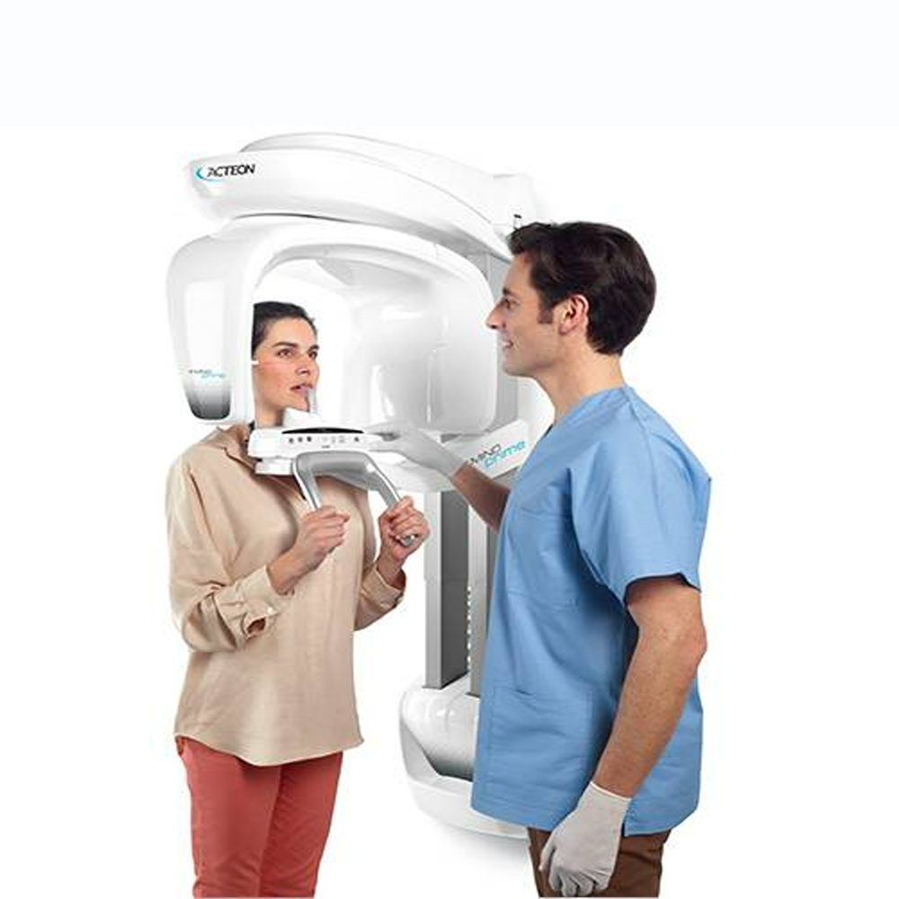
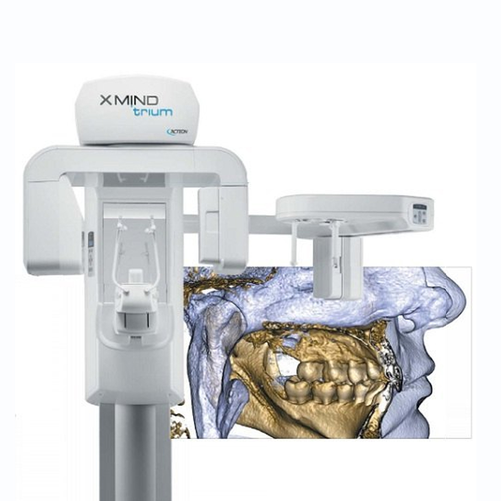
3D Dental Cone Beam Computed Tomography (CBCT) is an advanced imaging technology used in dentistry to obtain highly detailed three-dimensional images of the dental and maxillofacial structures. Unlike traditional dental X-rays, which provide two-dimensional images, CBCT offers a comprehensive view of the teeth, jawbone, temporomandibular joints, and surrounding soft tissues in a single scan.
CBCT scanners utilize a cone-shaped X-ray beam and a specialized detector to capture multiple images from different angles around the patient’s head. These images are then reconstructed into a three-dimensional volume using computer software, allowing dentists and oral surgeons to visualize the dental anatomy from various perspectives.
One of the key advantages of CBCT imaging is its ability to provide precise anatomical information, which is essential for accurate diagnosis and treatment planning in various dental procedures. For example, CBCT scans are commonly used in implant dentistry to assess bone quality and quantity, determine optimal implant placement sites, and visualize the proximity of vital structures such as nerves and sinuses.
In addition to implant planning, CBCT imaging is valuable in orthodontics for evaluating dental and skeletal relationships, assessing airway dimensions, and identifying impacted teeth. It is also used in endodontics to detect root canal anomalies and evaluate the morphology of root canal systems.
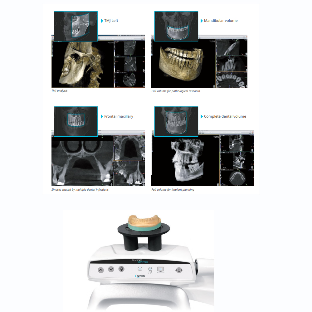
Furthermore, CBCT plays a crucial role in the diagnosis and treatment of temporomandibular joint (TMJ) disorders, as it allows for the assessment of joint morphology, condylar position, and disc displacement.
Despite its numerous benefits, CBCT imaging involves exposure to ionizing radiation, albeit at a significantly lower dose compared to medical CT scans. Therefore, careful consideration of the risks and benefits is essential, and CBCT scans should only be performed when necessary and justified.
Overall, 3D Dental Cone Beam Computed Tomography (CBCT) is a valuable tool in modern dentistry, offering detailed and comprehensive imaging for improved diagnosis, treatment planning, and patient care.
What is 3D Dental Cone Beam Computed Tomography (CBCT) and How Does It Work?

3D Dental Cone Beam Computed Tomography (CBCT) is a specialized imaging technique used in dentistry to produce detailed three-dimensional images of the teeth, jaw, and surrounding structures. It works by capturing multiple X-ray images from different angles around the head, which are then reconstructed by a computer to create a comprehensive 3D model of the oral and maxillofacial region, allowing for precise diagnosis and treatment planning in various dental procedures.
Basic Principles and Principles of 3D Dental CBCT:
3D Dental Cone Beam Computed Tomography (CBCT) operates on the same basic principles as traditional CT imaging, utilizing X-rays to produce cross-sectional images of the dental and jaw structures. However, CBCT is specifically designed for dental applications, offering higher resolution and lower radiation exposure compared to conventional CT scans.
How is the 3D Imaging of Dental and Jaw Structures Achieved?:
CBCT imaging involves the use of a cone-shaped X-ray beam that rotates around the patient’s head to capture multiple images from various angles. These images are then reconstructed by specialized software to create a detailed three-dimensional representation of the teeth, jawbones, nerve pathways, and surrounding soft tissues. This provides dentists with valuable information about the anatomy and condition of the oral and maxillofacial region.
The Role and Importance of 3D Dental CBCT in Dentistry:
3D Dental CBCT plays a crucial role in modern dentistry by providing detailed and accurate imaging of dental and jaw structures, enabling dentists to make more informed diagnoses and treatment plans. It is used in various dental procedures, including implant placement, orthodontic treatment planning, endodontic therapy, and oral surgery. CBCT imaging offers numerous benefits, such as improved treatment outcomes, reduced risk of complications, and enhanced patient care, making it an essential tool in contemporary dental practice.
Contributions of 3D Dental CBCT to Detailed Evaluation of Dental and Jaw Structures
3D Dental Cone Beam Computed Tomography (CBCT) significantly contributes to the detailed evaluation of dental and jaw structures by providing high-resolution, three-dimensional images of the oral and maxillofacial region. This technology allows for precise visualization of anatomical structures, including teeth, roots, bone, nerves, and soft tissues, from multiple angles and perspectives. With 3D CBCT imaging, dental professionals can accurately assess the morphology, size, shape, and position of teeth and roots, detect abnormalities such as cysts, tumors, or fractures, and evaluate bone quality and density. This comprehensive evaluation aids in treatment planning for various dental procedures, including implant placement, endodontic therapy, orthodontic treatment, and oral surgery, ultimately leading to improved clinical outcomes and patient care.
Applications and Uses of 3D Dental CBCT in Dentistry
The applications and uses of 3D Dental Cone Beam Computed Tomography (CBCT) in dentistry are extensive and diverse, encompassing various diagnostic, treatment planning, and therapeutic purposes.
Implant Dentistry: CBCT plays a crucial role in implant dentistry by providing detailed anatomical information about the alveolar bone, adjacent structures, and anatomical limitations, facilitating precise implant planning and placement for optimal aesthetics and function.
Endodontics: In endodontics, CBCT imaging aids in the diagnosis and treatment planning of complex root canal anatomy, periapical pathology, and root fractures, leading to more successful outcomes in root canal therapy.
Orthodontics: CBCT helps orthodontists assess skeletal relationships, identify impacted teeth, evaluate airway dimensions, and analyze temporomandibular joint (TMJ) morphology, assisting in comprehensive orthodontic diagnosis and treatment planning.
Oral and Maxillofacial Surgery: CBCT is indispensable in oral and maxillofacial surgery for preoperative assessment of facial fractures, impacted teeth, TMJ disorders, planning orthognathic surgery, and evaluating bone quality and quantity for bone grafting procedures.
Temporomandibular Joint (TMJ) Analysis: CBCT allows for accurate assessment of TMJ morphology, condylar position, disc displacement, and bony changes, aiding in the diagnosis and management of TMJ disorders.
Periodontics: CBCT assists periodontists in assessing bone levels, bone defects, furcation involvement, and root morphology, facilitating precise diagnosis and treatment planning for periodontal diseases and implant placement.
Pediatric Dentistry: CBCT imaging is beneficial in pediatric dentistry for evaluating dental anomalies, assessing dental eruption patterns, diagnosing dental trauma, and planning orthodontic treatment in growing children.
Forensic Dentistry: CBCT provides detailed three-dimensional images that are valuable in forensic odontology for identifying dental characteristics, assessing dental anomalies, and aiding in human identification in cases of mass disasters or forensic investigations.
Overall, the diverse applications of 3D Dental CBCT contribute to enhanced diagnostic accuracy, treatment precision, and patient care across various dental specialties.
Clinical Applications of 3D Dental CBCT Scanning
The clinical applications of 3D Dental Cone Beam Computed Tomography (CBCT) scanning span across various dental specialties, offering invaluable diagnostic and treatment planning capabilities. Some key clinical applications include:
Implant Dentistry: CBCT is extensively used in implant dentistry for preoperative assessment of bone volume, density, and quality, aiding in precise implant planning, placement, and evaluation of osseointegration.
Endodontics: CBCT imaging facilitates accurate diagnosis and treatment planning in endodontics by providing detailed three-dimensional images of tooth anatomy, periapical pathologies, root canal morphology, and complex root canal systems.
Orthodontics: CBCT plays a vital role in orthodontic treatment planning by providing detailed assessments of skeletal relationships, tooth angulation, impactions, airway dimensions, and temporomandibular joint (TMJ) morphology.
Oral and Maxillofacial Surgery: CBCT imaging is essential in oral and maxillofacial surgery for preoperative assessment of impacted teeth, facial fractures, TMJ disorders, cleft palate evaluation, orthognathic surgery planning, and evaluation of bone grafting sites.
Temporomandibular Joint (TMJ) Analysis: CBCT allows for comprehensive evaluation of TMJ morphology, condylar position, disc displacement, and bony changes, aiding in the diagnosis and management of TMJ disorders.
Periodontics: CBCT assists periodontists in assessing bone levels, bone defects, furcation involvement, and root morphology, facilitating precise diagnosis and treatment planning for periodontal diseases and dental implant placement.
Pediatric Dentistry: CBCT imaging is beneficial in pediatric dentistry for evaluating dental anomalies, assessing dental eruption patterns, diagnosing dental trauma, and planning orthodontic treatment in growing children.
Forensic Dentistry: CBCT provides detailed three-dimensional images that are valuable in forensic odontology for identifying dental characteristics, assessing dental anomalies, and aiding in human identification in cases of mass disasters or forensic investigations.
Overall, 3D Dental CBCT scanning has revolutionized clinical dentistry by offering advanced diagnostic capabilities, improving treatment outcomes, and enhancing patient care across a wide range of dental specialties.
The Role of 3D Dental CBCT in Planning and Placement of Implant Treatments
The role of 3D Dental Cone Beam Computed Tomography (CBCT) in planning and placement of implant treatments is paramount, offering detailed anatomical information crucial for successful outcomes. Here are key aspects of its role:
Preoperative Assessment: CBCT provides comprehensive preoperative assessment by offering detailed three-dimensional images of the alveolar bone, including bone density, volume, and morphology. This allows for precise evaluation of implant site anatomy, including bone quality, quantity, and proximity to vital structures like nerves and sinuses.
Implant Positioning and Orientation: CBCT aids in accurate implant positioning and orientation by visualizing bone dimensions and anatomical structures in three dimensions. It allows for virtual implant placement, enabling the clinician to select the optimal implant size, length, and angulation to achieve ideal implant placement relative to adjacent teeth and prosthetic considerations.
Surgical Guide Fabrication: CBCT images serve as the foundation for fabricating surgical guides, which are essential for precise implant placement. By superimposing virtual implant positions onto CBCT images, customized surgical guides can be designed to transfer the planned implant positions to the surgical site accurately, ensuring precise implant placement during surgery.
Bone Grafting and Augmentation: CBCT helps in assessing bone volume and morphology, guiding the need for bone grafting or augmentation procedures to enhance the implant site’s bone quality and quantity. It allows for accurate preoperative planning of bone grafting procedures and evaluation of graft integration postoperatively.
Risk Assessment and Complication Prevention: CBCT aids in identifying potential anatomical variations, such as sinus proximity, nerve canal deviation, or inadequate bone volume, which may increase the risk of surgical complications during implant placement. By identifying and addressing these factors preoperatively, CBCT contributes to minimizing surgical risks and preventing complications.
Patient Education and Informed Consent: CBCT images provide visual information to patients, helping them understand the proposed treatment plan, potential risks, and expected outcomes of implant placement. This facilitates informed consent by allowing patients to make educated decisions about their dental implant treatment.
Overall, 3D Dental CBCT plays a crucial role in planning and placement of implant treatments by providing detailed anatomical information, facilitating precise surgical planning, reducing surgical risks, and optimizing treatment outcomes in implant dentistry.
Advantages and Benefits of 3D Dental Cone Beam Computed Tomography (CBCT) include:
High-Resolution and Detailed Imaging: CBCT provides high-resolution three-dimensional images of dental and jaw structures, allowing for detailed evaluation of bone morphology, density, and anatomical landmarks. This detailed imaging enhances diagnostic accuracy and treatment planning precision.
Less Invasive Method: Compared to traditional imaging techniques like conventional radiography or medical CT scans, CBCT is less invasive for both patients and dentists. It requires lower radiation doses while providing comprehensive 3D imaging, reducing the potential risks associated with radiation exposure.
Efficient Treatment Planning: CBCT facilitates efficient treatment planning by providing comprehensive anatomical information in three dimensions. Dentists can accurately assess bone quality, volume, and morphology, leading to more precise implant placement, endodontic treatment planning, orthodontic assessment, and other dental procedures.
Improved Patient Care: The detailed imaging provided by CBCT enables dentists to offer personalized and tailored treatment plans based on each patient’s unique anatomical characteristics. This individualized approach improves patient care outcomes and enhances overall treatment satisfaction.
Enhanced Surgical Precision: CBCT aids in enhancing surgical precision by allowing dentists to visualize anatomical structures from multiple angles and perspectives. This enables precise preoperative planning and intraoperative guidance during dental implant placement, oral surgery, and other invasive procedures.
Minimized Radiation Exposure: While CBCT uses X-ray technology, it delivers lower radiation doses compared to conventional CT scans, making it a safer option for patients. Advanced imaging protocols and software optimization further minimize radiation exposure while maintaining diagnostic image quality.
Time and Cost Savings: CBCT streamlines the diagnostic process by providing comprehensive imaging in a single scan, reducing the need for multiple imaging modalities and appointments. This saves both time and costs for both patients and dental practices, leading to improved workflow efficiency.
Research and Education: CBCT has become an invaluable tool for dental research and education due to its ability to provide detailed three-dimensional images of dental and maxillofacial structures. It facilitates advanced research studies, treatment simulations, and educational demonstrations in various dental specialties.
Overall, 3D Dental CBCT offers numerous advantages and benefits, including high-resolution imaging, enhanced treatment planning, improved patient care, enhanced surgical precision, minimized radiation exposure, time and cost savings, and its utility in research and education within the field of dentistry.
Radiation exposure in dental cone beam computed tomography (CBCT) is a concern due to the use of X-ray technology. However, several safety measures are implemented to minimize radiation exposure and ensure patient safety:
Optimized Imaging Protocols: CBCT systems are equipped with optimized imaging protocols that use the lowest radiation dose necessary to produce diagnostic images. These protocols consider factors such as patient age, size, and the specific imaging requirements to minimize radiation exposure while maintaining image quality.
Beam Collimation and Limitation: CBCT systems employ beam collimation techniques to restrict the X-ray beam to the area of interest, minimizing unnecessary radiation exposure to surrounding tissues. Additionally, the field of view (FOV) can be adjusted to limit the imaging area to the specific region of interest, further reducing radiation exposure.
Pulsed X-ray Technology: Some CBCT systems utilize pulsed X-ray technology, where the X-ray beam is delivered in short bursts rather than continuously. This reduces overall radiation exposure while still obtaining diagnostic images.
Shielding and Lead Aprons: Lead aprons and thyroid collars are commonly used to shield sensitive organs from radiation during CBCT scanning. Lead shields may also be used to cover areas not included in the imaging field, further reducing scatter radiation.
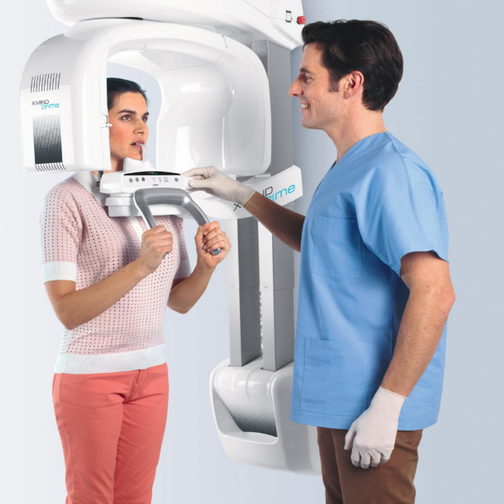
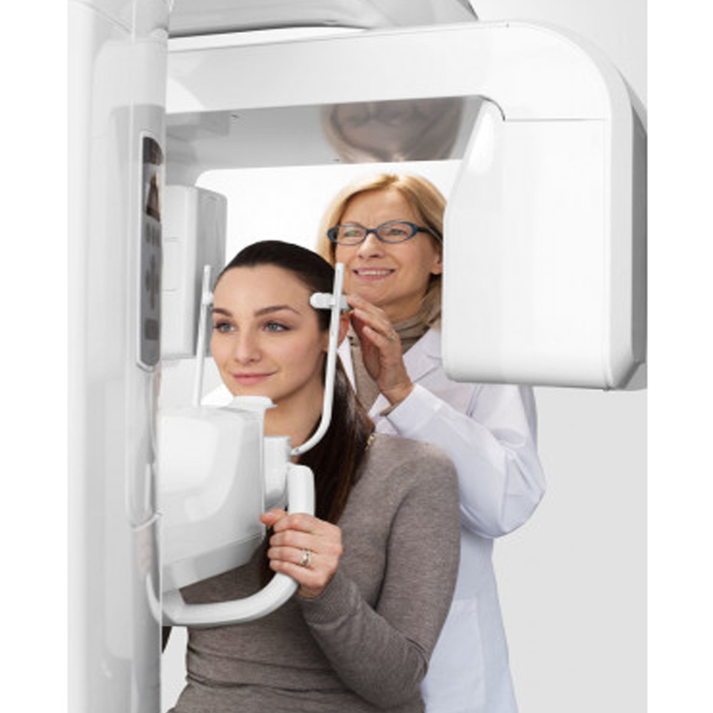
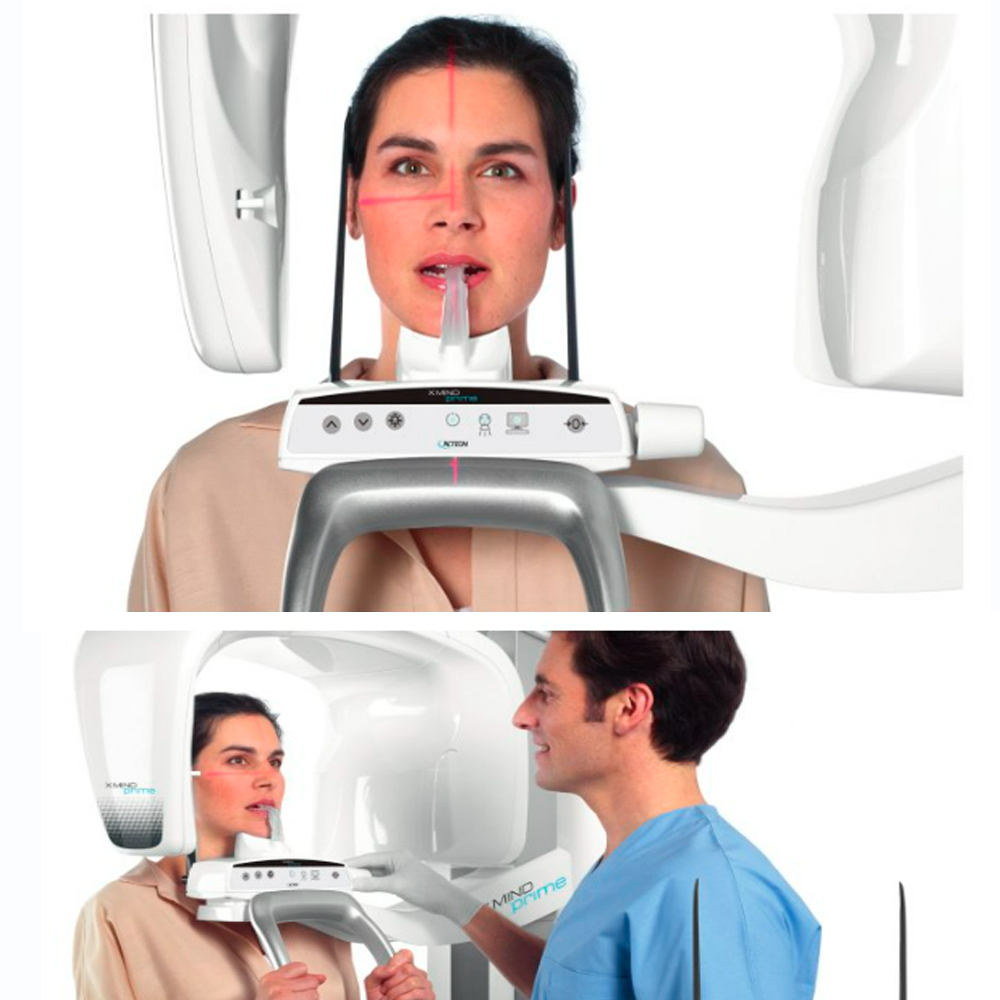
Patient Positioning and Immobilization: Proper patient positioning and immobilization techniques are essential to ensure accurate imaging and minimize the need for repeat scans. This reduces the overall number of exposures and radiation dose received by the patient.
ALARA Principle: The ALARA (As Low As Reasonably Achievable) principle guides CBCT imaging practices, emphasizing the importance of minimizing radiation exposure while obtaining diagnostic images of adequate quality. This principle encourages clinicians to use the lowest radiation dose necessary for the clinical task at hand.
Operator Training and Certification: CBCT operators undergo specialized training and certification to ensure they understand radiation safety principles, proper equipment operation, and imaging protocols. This helps minimize errors and optimize imaging practices to reduce radiation exposure.
Patient Education: Patients are informed about the risks and benefits of CBCT imaging, including radiation exposure, before undergoing the procedure. Informed consent is obtained, and patients are encouraged to ask questions and express any concerns they may have regarding radiation safety.
By implementing these radiation safety measures, dental professionals can effectively minimize radiation exposure during CBCT imaging while ensuring accurate diagnostic information for treatment planning.
Precautions for Patient Safety During Scanning:
- Patient Positioning: Ensuring proper patient positioning during scanning is crucial to obtain accurate images and minimize radiation exposure. Patients should be positioned comfortably and securely to prevent movement during the scan.
- Immobilization Devices: The use of immobilization devices, such as bite blocks or headrests, helps stabilize the patient’s position and reduce motion artifacts, improving image quality while minimizing the need for repeat scans.
- Radiation Shielding: Lead aprons and thyroid collars are commonly used to shield sensitive areas from radiation exposure during CBCT scanning. Additionally, lead shields may be used to protect areas not included in the imaging field.
- Beam Collimation: CBCT systems utilize beam collimation techniques to restrict the X-ray beam to the area of interest, minimizing unnecessary radiation exposure to surrounding tissues.
- Optimized Imaging Protocols: Employing optimized imaging protocols tailored to the specific clinical task and patient characteristics helps minimize radiation dose while ensuring diagnostic image quality.
- Patient Education: Providing patients with information about the CBCT procedure, including radiation exposure risks and safety measures, allows them to make informed decisions and feel more comfortable during the scan.
Evaluation and Interpretation of 3D Dental CBCT Images:
- Image Reconstruction: CBCT images are reconstructed using specialized software, and careful attention should be paid to ensure proper image reconstruction settings are selected to optimize image quality and diagnostic information.
- Anatomical Landmarks: Identifying anatomical landmarks and structures within the CBCT images is essential for accurate interpretation and diagnosis. Clinicians should be familiar with dental anatomy and pathology to interpret images effectively.
- Pathological Findings: CBCT images may reveal various pathological findings, such as dental caries, periodontal disease, or impacted teeth. Thorough evaluation of these findings is necessary for accurate diagnosis and treatment planning.
- Image Artefacts: Awareness of common image artefacts in CBCT imaging, such as streaking or beam hardening artefacts, is important to distinguish them from true anatomical structures and avoid misinterpretation.
- Consultation with Specialists: In complex cases or when interpreting findings outside of their expertise, clinicians may consult with radiologists or other specialists to ensure accurate diagnosis and appropriate treatment planning.
Understanding Image Results and Sharing with the Doctor:
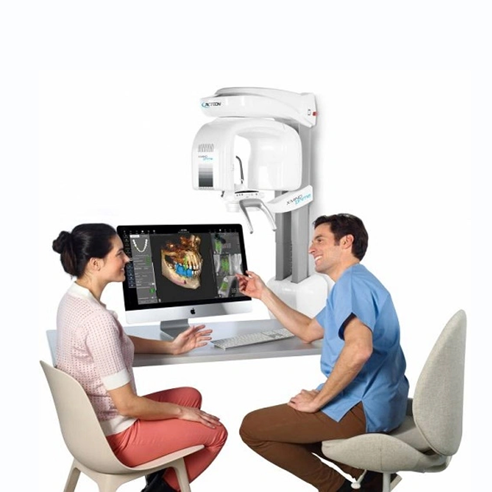
- Patient Communication: After interpreting CBCT images, clinicians should communicate the findings clearly and comprehensively to patients, using layman’s terms to ensure understanding.
- Treatment Options: Explaining the implications of CBCT findings on treatment options and discussing potential treatment plans with patients allows for informed decision-making and patient involvement in the treatment process.
- Collaboration with Referring Doctors: If the CBCT scan was ordered by a referring doctor, clear and concise communication of the imaging findings and their implications facilitates collaboration between clinicians and ensures coordinated patient care.
Use of 3D Dental CBCT Images in the Treatment Process:
- Treatment Planning: 3D dental CBCT images provide detailed anatomical information that aids in treatment planning for various dental procedures, including implant placement, orthodontic treatment, endodontic therapy, and oral surgeries.
- Precise Implant Placement: CBCT images allow for precise assessment of bone quality, quantity, and location, facilitating optimal implant placement and reducing the risk of complications.
- Orthodontic Treatment: CBCT imaging enables orthodontists to visualize tooth roots, impacted teeth, and the surrounding bone structure in three dimensions, enhancing treatment planning accuracy and efficiency.
- Endodontic Therapy: CBCT scans aid endodontists in diagnosing complex root canal anatomy, identifying periapical lesions, and assessing the extent of root resorption, leading to more successful endodontic outcomes.
Improvements in 3D Dental CBCT Imaging Techniques and Future Potential:
- Advanced Image Reconstruction Algorithms: Ongoing advancements in image reconstruction algorithms enhance image quality, reducing artifacts and improving diagnostic accuracy.
- Reduced Radiation Dose: Research efforts focus on developing low-dose CBCT protocols without compromising image quality, ensuring patient safety while maintaining diagnostic efficacy.
- Integration with CAD/CAM Technology: Integration of CBCT imaging with computer-aided design/computer-aided manufacturing (CAD/CAM) technology facilitates virtual treatment planning and fabrication of custom dental prostheses with precise fit and esthetics.
- Artificial Intelligence (AI) Applications: AI-driven algorithms hold promise for automated image analysis, aiding in faster and more accurate diagnosis of dental conditions and treatment planning.
Patient Rights and Information About 3D Dental CBCT Scanning:
- Informed Consent: Patients should receive comprehensive information about the purpose of the CBCT scan, associated risks, benefits, and alternative imaging modalities before providing informed consent for the procedure.
- Radiation Safety: Patients have the right to know about radiation exposure risks associated with CBCT imaging and the measures taken to minimize radiation dose, such as beam collimation and shielding.
- Privacy and Confidentiality: Healthcare providers must adhere to strict privacy regulations and ensure patient data confidentiality when acquiring, storing, and sharing CBCT images, in compliance with HIPAA and other applicable laws.
- Access to Imaging Results: Patients have the right to access their CBCT imaging results and should be provided with explanations and interpretations in a clear and understandable manner by their dental healthcare providers.
- Patient Preferences: Dentists should respect patient preferences regarding the use of CBCT imaging and involve them in decision-making processes related to treatment planning and imaging options.
By upholding patient rights and providing comprehensive information, dental healthcare providers ensure patient autonomy, safety, and satisfaction throughout the CBCT scanning process.
Privacy and Data Protection:
- Confidentiality: Dental healthcare providers must ensure the confidentiality of patient data collected during 3D dental CBCT scanning, adhering to strict privacy regulations such as the Health Insurance Portability and Accountability Act (HIPAA) in the United States and similar laws worldwide.
- Secure Data Storage: Patient CBCT images and related data should be securely stored in encrypted digital formats and accessible only to authorized personnel involved in patient care, with robust measures in place to prevent unauthorized access or data breaches.
- Data Transmission: When transmitting CBCT images electronically for consultation or referral purposes, secure channels such as encrypted emails or secure file transfer protocols (SFTP) should be used to safeguard patient data during transit.
- Patient Consent: Dental healthcare providers should obtain explicit consent from patients before sharing their CBCT images or related data with third parties for educational, research, or other purposes, ensuring transparency and patient autonomy.
- Data Retention Policies: Healthcare facilities should establish clear policies regarding the retention and disposal of patient CBCT images and associated data, ensuring compliance with legal requirements and protecting patient privacy.
- Regular Security Audits: Dental practices should conduct regular security audits and risk assessments to identify vulnerabilities in their data protection measures and implement necessary safeguards to mitigate potential risks of data breaches or unauthorized access.
- Staff Training: All staff members handling patient CBCT images and data should undergo training on data protection protocols, including confidentiality agreements, secure data handling procedures, and awareness of potential cybersecurity threats.
- Vendor Compliance: Dental practices utilizing third-party vendors or cloud-based services for CBCT image storage or data management should ensure that these vendors comply with stringent data protection standards and adhere to relevant regulatory requirements.
- Patient Rights: Patients have the right to request access to their CBCT images and related data, as well as the right to request corrections or amendments to inaccuracies in their records, in accordance with applicable data protection laws and regulations.
By prioritizing privacy and implementing robust data protection measures, dental healthcare providers uphold patient trust, ensure compliance with regulatory requirements, and safeguard patient confidentiality throughout the 3D dental CBCT scanning process.
Patient Rights and Preferences During the Scanning Process:
- Informed Consent: Patients undergoing 3D dental CBCT scanning have the right to receive comprehensive information about the procedure, including its purpose, benefits, potential risks, and alternatives, enabling them to make informed decisions about their healthcare.
- Privacy and Dignity: Patients should be treated with respect for their privacy and dignity during the scanning process, with measures in place to ensure confidentiality and modesty, such as providing appropriate attire and maintaining a private environment.
- Comfort and Safety: Dental healthcare providers should prioritize patient comfort and safety during CBCT scanning, addressing any concerns or discomfort experienced by patients and taking necessary precautions to minimize radiation exposure and ensure the highest standard of care.
- Patient Preferences: Healthcare providers should respect patient preferences regarding the timing, location, and other aspects of the scanning process whenever feasible, accommodating individual needs and preferences to enhance the patient experience and promote satisfaction with care.
- Right to Refuse: Patients have the right to refuse or withdraw consent for 3D dental CBCT scanning at any time, without fear of coercion or discrimination, with healthcare providers respecting their autonomy and providing alternative options for diagnosis or treatment.
Future and Development of Imaging Techniques in Dentistry:
- Technological Advancements: The future of imaging techniques in dentistry is characterized by ongoing technological advancements, including innovations in 3D dental CBCT technology, such as improved image resolution, faster scanning times, and enhanced diagnostic capabilities.
- Integration of Artificial Intelligence (AI): AI-driven algorithms and machine learning techniques are increasingly being integrated into dental imaging systems, enabling automated image analysis, computer-aided diagnosis, and personalized treatment planning based on CBCT data.
- Multimodal Imaging: Future developments may involve the integration of 3D dental CBCT with other imaging modalities, such as intraoral scanners, digital impressions, and photogrammetry, to provide comprehensive diagnostic information and enhance treatment outcomes.
- Point-of-Care Imaging: Advancements in portable and handheld CBCT devices may enable point-of-care imaging in dental practices, facilitating immediate diagnosis and treatment planning at chairside, thereby improving efficiency and patient convenience.
- Customized Treatment Planning: Future imaging techniques aim to facilitate customized treatment planning in dentistry by providing detailed anatomical information, predictive analytics, and virtual simulations based on 3D dental CBCT data, allowing for more precise and personalized interventions.
Advancements in 3D Dental CBCT Technology and Future Directions:
- Miniaturization and Portability: Future developments may focus on miniaturizing CBCT systems and enhancing their portability, making them more accessible for use in diverse clinical settings, including mobile dental units, community health centers, and remote areas.
- Enhanced Imaging Capabilities: Ongoing advancements in CBCT technology aim to improve image quality, resolution, and contrast enhancement, enabling better visualization of dental and craniofacial structures and enhancing diagnostic accuracy for various dental conditions.
- Reduced Radiation Exposure: Future CBCT systems may incorporate innovative dose reduction techniques, such as iterative reconstruction algorithms, optimized exposure parameters, and low-dose imaging protocols, to minimize radiation exposure while maintaining diagnostic image quality.
- Integration with CAD/CAM Systems: Integration of 3D dental CBCT with computer-aided design and computer-aided manufacturing (CAD/CAM) systems allows for seamless transfer of digital impressions, virtual treatment planning, and fabrication of custom dental prostheses, facilitating more efficient and precise restorative procedures.
- Interdisciplinary Applications: Future directions in 3D dental CBCT technology include expanding its interdisciplinary applications beyond dentistry to fields such as oral and maxillofacial surgery, orthodontics, implantology, temporomandibular joint (TMJ) imaging, and forensic dentistry, where detailed anatomical information is crucial for accurate diagnosis and treatment planning.
By addressing patient rights and preferences, anticipating future trends in imaging techniques, and embracing technological advancements in 3D dental CBCT technology, dental healthcare providers can enhance patient care, improve diagnostic accuracy, and optimize treatment outcomes in the evolving landscape of modern dentistry.
Comparison with Other Imaging Techniques: 3D dental CBCT offers several advantages over traditional 2D imaging techniques such as panoramic radiography and intraoral radiography. Unlike 2D images, CBCT provides volumetric data, allowing for accurate assessment of dental and maxillofacial structures in three dimensions. This comprehensive view enables better visualization of anatomical structures, improved assessment of bone quality and density, and more precise evaluation of dental pathologies and abnormalities. Additionally, CBCT imaging reduces the need for multiple exposures and offers lower radiation doses compared to conventional CT scans, making it a safer alternative for patients.
Future Perspectives: The future of 3D dental CBCT imaging holds significant potential for further advancements and innovations. Some potential future developments include:
Improved Image Resolution: Continued advancements in CBCT technology may lead to enhanced image resolution, allowing for better visualization of smaller anatomical structures and finer details.
Integration with Artificial Intelligence (AI): Integration of AI algorithms into CBCT software may enable automated image analysis, facilitating faster and more accurate diagnosis of dental conditions and treatment planning.
Functional Imaging: Future CBCT systems may incorporate functional imaging capabilities, such as dynamic imaging or real-time tracking of jaw movements during mastication, which could provide valuable insights for orthodontic and prosthodontic treatment planning.
Portable and Point-of-Care Devices: Development of compact and portable CBCT devices may enable their use in point-of-care settings, expanding access to advanced imaging technologies in dental clinics, emergency rooms, and remote healthcare facilities.
Multimodal Imaging Integration: Integration of CBCT with other imaging modalities, such as optical coherence tomography (OCT) or ultrasound, may provide complementary information and enhance diagnostic accuracy for certain dental conditions.
Regulatory Standards and Guidelines: Continued development and refinement of regulatory standards and guidelines for CBCT imaging will be essential to ensure consistent image quality, patient safety, and ethical use of this technology.
Overall, the future of 3D dental CBCT imaging is promising, with ongoing advancements expected to further improve diagnostic capabilities, treatment outcomes, and patient care in the field of dentistry.
Frequently Asked Questions

Get a Free Second Opinion

I consent to Burtom Health Group using my aforesaid personal data for the purposes described in this notice and understand that I can withdraw my consent at any time by sending a request to info@burtom.com.

