Burtom ‣ Technologies ‣ Digital Mammography (3D Tomosynthesis)
Language: 🇬🇧 English | 🇹🇷 Türkçe
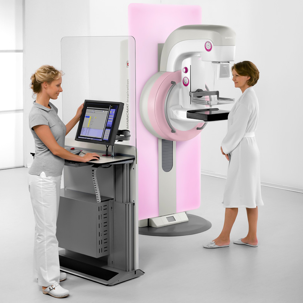
Digital Mammography (3D Tomosynthesis) Overview

 Digital mammography, also known as 3D tomosynthesis, represents a significant advancement in breast imaging technology, offering enhanced capabilities for breast cancer detection and diagnosis. Unlike conventional 2D mammography, which captures a single image of the breast, digital mammography utilizes multiple low-dose X-ray images taken from different angles to reconstruct a three-dimensional view of the breast tissue. This allows radiologists to examine the breast in thin slices, improving their ability to detect abnormalities such as tumors or masses that may be obscured in traditional mammograms.
One of the key benefits of digital mammography is its ability to reduce the impact of overlapping breast tissue, which can sometimes obscure abnormalities and lead to false-positive or false-negative results in 2D mammography. By capturing images from multiple angles and reconstructing them into a three-dimensional image, digital mammography provides clearer and more detailed images, leading to improved accuracy in breast cancer detection.
Digital mammography is particularly useful in imaging dense breast tissue, which is common among younger women and can make it challenging to detect abnormalities with traditional mammography. The ability to visualize breast tissue in thin slices helps radiologists identify subtle changes and detect abnormalities earlier, potentially leading to earlier diagnosis and more effective treatment.
Furthermore, digital mammography offers other advantages such as shorter examination times, reduced radiation exposure compared to film-based mammography, and the ability to store and transmit images electronically for easier access and sharing among healthcare providers. As technology continues to advance, digital mammography systems are also evolving to incorporate features like computer-aided detection (CAD) software, which can assist radiologists in interpreting images and identifying potential areas of concern.
In summary, digital mammography, or 3D tomosynthesis, represents a significant advancement in breast imaging technology, offering improved accuracy, enhanced visualization of breast tissue, and greater potential for early detection of breast cancer compared to traditional 2D mammography. Its ability to provide clearer and more detailed images makes it an invaluable tool in the fight against breast cancer, contributing to more effective screening, diagnosis, and treatment planning.


Digital Mammography and 3D Tomosynthesis: What are They and How Do They Work?
Digital mammography and 3D tomosynthesis are advanced imaging techniques used for the early detection and monitoring of breast cancer. Digital mammography is a screening method that digitally images breast tissue, while 3D tomosynthesis is an advanced technique used to obtain a detailed three-dimensional image of breast tissue. These methods provide more accurate results in breast cancer screening and are crucial tools for early diagnosis. Both digital mammography and 3D tomosynthesis are recognized as reliable and effective tools for breast cancer diagnosis and monitoring.
Basic Principles and Principles of Digital Mammography and 3D Tomosynthesis
Digital mammography utilizes digital detectors to capture X-ray images of breast tissue, offering advantages such as immediate image availability and the ability to manipulate image contrast and brightness. On the other hand, 3D tomosynthesis involves capturing multiple X-ray images of the breast from various angles and reconstructing them into a three-dimensional image, providing improved visualization of breast structures and lesions.
Evolution of Breast Imaging Techniques and Digital Transformation
The field of breast imaging has evolved significantly over the years, transitioning from traditional film-screen mammography to digital mammography and now to 3D tomosynthesis. This evolution has been driven by advancements in technology, leading to improved image quality, enhanced diagnostic accuracy, and reduced radiation exposure for patients.
Applications and Importance of Digital Mammography and 3D Tomosynthesis
Digital mammography and 3D tomosynthesis play crucial roles in breast cancer screening, diagnosis, and monitoring. They enable earlier detection of breast abnormalities, including tumors and microcalcifications, leading to improved patient outcomes and reduced mortality rates. Additionally, these advanced imaging techniques allow for more accurate lesion localization, leading to better surgical planning and treatment outcomes for patients with breast cancer.
Role and Contributions of Digital Imaging
Digital imaging technologies, including digital mammography and 3D tomosynthesis, have revolutionized breast cancer screening and diagnosis. By providing high-resolution images with enhanced contrast and detail, digital imaging techniques enable healthcare professionals to detect abnormalities in breast tissue more accurately and at earlier stages, contributing to improved patient outcomes and survival rates.
Digital Mammography and 3D Tomosynthesis Technology and Working Principle
Digital mammography utilizes digital detectors to capture X-ray images of breast tissue, offering advantages such as immediate image availability, improved image quality, and the ability to manipulate image contrast and brightness. On the other hand, 3D tomosynthesis involves capturing multiple X-ray images of the breast from various angles and reconstructing them into a three-dimensional image, providing enhanced visualization of breast structures and abnormalities.
Digital Mammography and 3D Tomosynthesis System and Basic Components
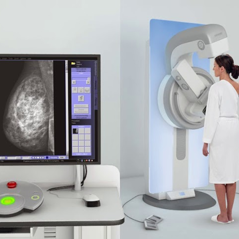
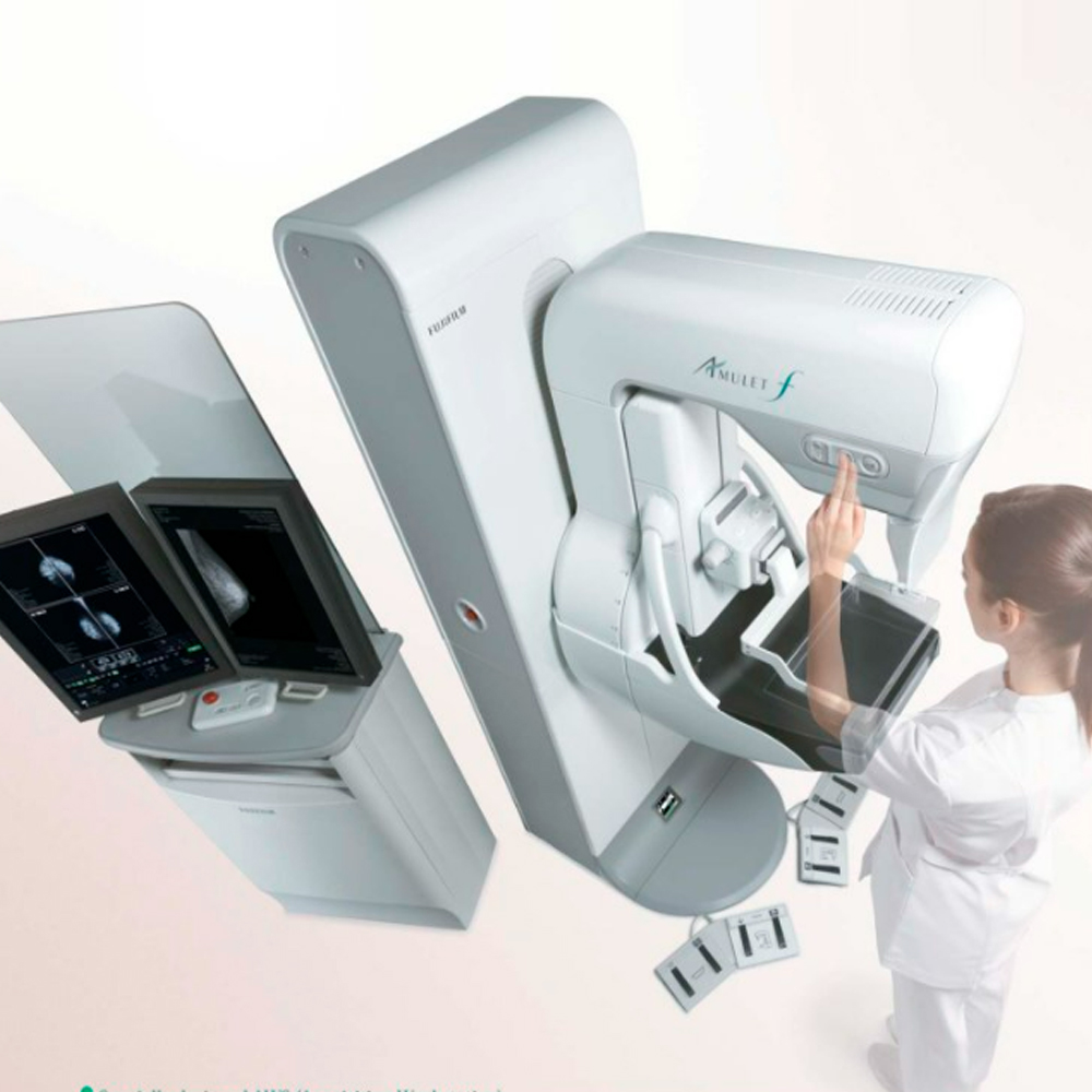
The digital mammography and 3D tomosynthesis systems consist of several key components, including X-ray tubes, detectors, image processing software, and display monitors. These components work together to capture, process, and display high-quality images of the breast, allowing healthcare professionals to accurately evaluate breast tissue and detect abnormalities with greater precision and efficiency.
Image Acquisition and Processing
Image acquisition in digital mammography and 3D tomosynthesis involves the use of X-ray technology to capture images of the breast tissue. These images are then processed using advanced digital imaging techniques to enhance contrast, reduce noise, and optimize image quality. Digital processing allows for immediate image availability and enables healthcare professionals to manipulate image parameters for improved visualization and analysis.
Clinical Applications of Digital Mammography and 3D Tomosynthesis
Digital mammography and 3D tomosynthesis play a crucial role in breast cancer screening, diagnosis, and treatment planning. They are widely used in clinical practice for routine mammographic screening, as well as for evaluating breast abnormalities detected on physical examination or other imaging modalities. Additionally, 3D tomosynthesis provides enhanced visualization of breast structures, improving the detection of breast lesions and reducing false-positive findings compared to traditional mammography.
Detailed Evaluation of Breast Tissue and Localization Determination
Digital mammography and 3D tomosynthesis enable detailed evaluation of breast tissue architecture and pathology, allowing healthcare professionals to accurately localize and characterize abnormalities such as masses, microcalcifications, and architectural distortions. This detailed evaluation aids in the diagnosis and staging of breast cancer, as well as in treatment planning and monitoring. By providing three-dimensional images of the breast, 3D tomosynthesis enhances localization accuracy and facilitates targeted biopsies, improving patient care and outcomes.
Advantages and Benefits of Digital Mammography and 3D Tomosynthesis
Digital mammography and 3D tomosynthesis offer several advantages over traditional film-screen mammography, providing high-resolution and detailed imaging of the breast tissue.
High Resolution and Detailed Imaging
Digital mammography and 3D tomosynthesis produce high-resolution images that allow for detailed visualization of breast structures, including subtle abnormalities that may not be visible on conventional mammograms. This improved imaging capability enhances the detection of breast lesions and abnormalities, enabling earlier diagnosis and more accurate characterization of breast pathology.
Precise Diagnosis with 3D Imaging and Fewer False Positive Results
3D tomosynthesis, in particular, allows for precise three-dimensional imaging of the breast, reducing the likelihood of false-positive findings compared to traditional two-dimensional mammography. By providing multiple images of the breast from different angles, 3D tomosynthesis improves the visualization of breast tissue and helps to distinguish between benign and malignant lesions, leading to more accurate diagnoses and fewer unnecessary callbacks for additional imaging or biopsies. This reduces patient anxiety and healthcare costs while improving overall diagnostic accuracy and patient outcomes.
Radiation Exposure and Safety Measures
Digital mammography and 3D tomosynthesis involve exposure to ionizing radiation, albeit at low doses considered safe for routine screening and diagnostic purposes. Nevertheless, measures are taken to minimize radiation exposure and ensure patient safety during scanning.
Precautions Taken for Patient Safety During Scanning
Healthcare providers follow strict protocols to minimize radiation exposure during digital mammography and 3D tomosynthesis procedures. This includes:
Optimizing Equipment Settings: Technicians adjust equipment settings to use the lowest possible radiation dose while still obtaining high-quality images.
Lead Aprons and Shields: Patients wear lead aprons or shields to protect sensitive organs from radiation exposure during imaging.
Collimation: Collimation techniques are employed to restrict the x-ray beam to the area of interest, reducing unnecessary radiation exposure to surrounding tissues.
Proper Positioning: Ensuring proper positioning of the patient and breast compression helps optimize image quality and minimize the need for repeat exposures.
Limiting Exposure Time: Technicians minimize the duration of exposure to ionizing radiation by efficiently performing the imaging procedure.
Evaluation and Interpretation of Digital Mammography and 3D Tomosynthesis Images
Following image acquisition, radiologists carefully evaluate and interpret digital mammography and 3D tomosynthesis images to detect and characterize abnormalities in breast tissue. This process involves:
Image Quality Assessment: Radiologists assess the quality of acquired images to ensure optimal visualization of breast structures and any potential abnormalities.
Lesion Detection: Radiologists meticulously examine digital mammography and 3D tomosynthesis images to identify suspicious lesions, such as masses, microcalcifications, or architectural distortions.
Characterization of Abnormalities: Radiologists characterize detected abnormalities based on their size, shape, margins, and other imaging features to determine the likelihood of malignancy and guide further diagnostic workup or intervention.
Comparative Analysis: Radiologists may compare current images with prior mammograms or imaging studies to assess changes over time and evaluate the progression or stability of detected abnormalities.
Reporting: Radiologists provide detailed reports of their findings, including descriptions of detected abnormalities, their characteristics, and recommendations for additional imaging, biopsy, or follow-up based on established diagnostic criteria and guidelines.
Understanding Image Results and Sharing with the Doctor
Understanding digital mammography and 3D tomosynthesis image results is crucial for accurate diagnosis and effective patient management. Radiologists interpret the images and communicate their findings to the referring healthcare provider, typically a primary care physician or a specialist such as a breast surgeon or oncologist. They explain the significance of any detected abnormalities, discuss potential implications for the patient’s health, and collaborate with the healthcare team to determine appropriate next steps, including additional imaging, biopsy, or referral to a specialist.
Use and Monitoring of Images in the Treatment Process
Digital mammography and 3D tomosynthesis images play a vital role in guiding treatment decisions and monitoring patient outcomes throughout the treatment process. Healthcare providers use these images to plan and monitor various interventions, such as surgical procedures, radiation therapy, or chemotherapy, aimed at managing breast cancer or other breast conditions. Additionally, digital mammography and 3D tomosynthesis images serve as valuable tools for assessing treatment response, detecting recurrent disease, and evaluating long-term outcomes, allowing for timely adjustments to treatment strategies as needed.
Technological Developments and Innovations in Digital Mammography and 3D Tomosynthesis
Advancements in digital mammography and 3D tomosynthesis technology continue to enhance breast imaging capabilities, leading to improved diagnostic accuracy, enhanced patient comfort, and reduced radiation exposure. Key technological developments and innovations in this field include:
Improved Image Resolution: Ongoing advancements in detector technology and image processing algorithms result in higher image resolution, enabling better visualization of breast tissue structures and abnormalities.
Artificial Intelligence (AI) Integration: Integration of AI algorithms into digital mammography and 3D tomosynthesis systems enhances image interpretation by assisting radiologists in lesion detection, characterization, and risk assessment, thereby improving diagnostic accuracy and efficiency.
Iterative Reconstruction Techniques: Iterative reconstruction algorithms optimize image quality while minimizing radiation dose, allowing for high-quality imaging with reduced patient exposure to ionizing radiation.
3D Printing for Surgical Planning: Incorporating 3D printing technology into digital mammography and tomosynthesis facilitates the creation of patient-specific anatomical models, aiding surgeons in preoperative planning and intraoperative navigation for breast-conserving surgeries and reconstructive procedures.
Advanced Visualization Tools: Innovative visualization tools, such as multiplanar reconstruction and 3D volume rendering, provide radiologists with comprehensive insights into breast anatomy and pathology, facilitating accurate interpretation and confident diagnosis.
These technological developments underscore the continuous evolution of digital mammography and 3D tomosynthesis, empowering healthcare providers with advanced imaging solutions for optimal breast care and improved patient outcomes.
Improvements in Imaging Techniques and Future Potential
Continual advancements in digital mammography and 3D tomosynthesis imaging techniques hold promise for enhancing breast cancer detection, diagnosis, and treatment monitoring. Future developments may focus on:
Enhanced Image Quality: Ongoing improvements in image resolution, contrast enhancement, and artifact reduction techniques aim to provide clearer and more detailed images for improved diagnostic accuracy.
Quantitative Imaging Biomarkers: Research into quantitative imaging biomarkers, such as breast density measurements and tissue stiffness assessments, may enable more precise risk stratification, early detection of abnormalities, and personalized treatment planning.
Functional Imaging Techniques: Emerging functional imaging modalities, including contrast-enhanced mammography and diffusion-weighted imaging, offer insights into tissue physiology and microstructural changes, potentially improving the characterization of breast lesions and treatment monitoring.
Integration with Molecular Imaging: Integration of digital mammography and 3D tomosynthesis with molecular imaging modalities, such as positron emission mammography (PEM) and molecular breast imaging (MBI), may enable comprehensive assessment of breast tissue morphology and molecular characteristics, facilitating personalized management strategies.
Artificial Intelligence (AI) Applications: Continued integration of AI algorithms into digital mammography and 3D tomosynthesis systems holds promise for automated lesion detection, computer-aided diagnosis, and personalized risk assessment, potentially improving workflow efficiency and diagnostic accuracy.
Patient Rights and Important Considerations Regarding Digital Mammography and 3D Tomosynthesis
Patients undergoing digital mammography and 3D tomosynthesis have rights and considerations that healthcare providers must prioritize, including:
Informed Consent: Patients have the right to receive comprehensive information about the benefits, risks, and alternatives of digital mammography and 3D tomosynthesis before providing informed consent for the procedure.
Privacy and Confidentiality: Healthcare providers must adhere to strict privacy and data protection regulations to safeguard patients’ sensitive health information collected during digital mammography and 3D tomosynthesis examinations.
Dignity and Respect: Patients have the right to be treated with dignity, respect, and sensitivity throughout the imaging process, with healthcare providers ensuring their comfort and privacy during the procedure.
Access to Results: Patients are entitled to timely access to their digital mammography and 3D tomosynthesis results, accompanied by clear explanations from their healthcare providers to aid in understanding and decision-making regarding further evaluation or treatment.
Patient Education: Healthcare providers should educate patients about the importance of regular breast cancer screening, the role of digital mammography and 3D tomosynthesis in early detection, and the significance of adhering to recommended screening guidelines based on individual risk factors.
Privacy and Data Protection
Protecting patient privacy and ensuring data security are paramount considerations in digital mammography and 3D tomosynthesis:
Secure Data Storage: Healthcare facilities must employ robust data encryption and secure storage measures to protect patients’ digital mammography and 3D tomosynthesis images and associated health information from unauthorized access or breaches.
Compliance with Regulations: Healthcare providers must adhere to stringent regulatory standards, such as the Health Insurance Portability and Accountability Act (HIPAA) in the United States, to maintain patient confidentiality and comply with privacy regulations governing medical imaging data.
Informed Consent for Data Sharing: Patients should be informed about any potential data sharing or research use of their digital mammography and 3D tomosynthesis images and have the opportunity to provide consent or opt-out based on their preferences.
Patient Rights and Preferences During the Scanning Process
During digital mammography and 3D tomosynthesis scanning, patients have certain rights and preferences that should be respected to ensure a positive and comfortable experience. These rights include:
Informed Consent: Patients have the right to receive clear and comprehensive information about the procedure, including its purpose, potential risks and benefits, and alternative options. They should provide informed consent before undergoing the imaging examination.
Privacy and Dignity: Patients have the right to privacy and dignity during the scanning process. Healthcare providers should ensure that the patient’s personal information is kept confidential, and modesty should be maintained throughout the procedure.
Comfort and Safety: Patients have the right to receive care in a safe and comfortable environment. Healthcare providers should take measures to minimize discomfort during the imaging procedure, such as providing appropriate positioning aids and addressing any concerns or anxieties the patient may have.
Respect for Preferences: Patients have the right to express their preferences regarding the scanning process, such as their preferred position during the examination or any specific accommodations they may require. Healthcare providers should respect these preferences whenever possible.
Future and Development of Breast Cancer Screening
The future of breast cancer screening holds promising advancements aimed at improving early detection and patient outcomes. Some key developments and future directions include:
Personalized Screening Guidelines: Advances in risk stratification models and genetic testing may lead to more personalized breast cancer screening guidelines tailored to individual risk profiles, optimizing the balance between benefits and harms of screening for different populations.
Integration of Artificial Intelligence (AI): AI-driven technologies, including machine learning algorithms and deep learning techniques, have the potential to enhance mammographic interpretation by improving lesion detection, characterization, and risk assessment, thereby reducing false positives and false negatives.
Multimodal Imaging Approaches: Combining digital mammography and 3D tomosynthesis with other imaging modalities, such as breast MRI or molecular breast imaging, may offer complementary information and improve diagnostic accuracy, particularly in women with dense breast tissue or high-risk factors.
Innovations in Image Acquisition: Advancements in imaging technology, such as photon-counting detectors and spectral imaging, hold promise for enhancing image quality, reducing radiation dose, and enabling novel imaging techniques for early cancer detection and characterization.
Precision Medicine and Targeted Therapies: Continued advancements in molecular profiling and targeted therapies may revolutionize breast cancer management, leading to more personalized treatment approaches based on the molecular characteristics of individual tumors.
Advancements in Digital Mammography and 3D Tomosynthesis Technology and Future Directions
The field of digital mammography and 3D tomosynthesis is continuously evolving, driven by technological innovations and research advancements. Some key advancements and future directions include:
Improved Image Quality: Ongoing research focuses on enhancing image resolution, contrast, and spatial resolution in digital mammography and 3D tomosynthesis to improve lesion detection and characterization, particularly in women with dense breast tissue.
Reduced Radiation Dose: Efforts are underway to develop novel imaging techniques and reconstruction algorithms that enable high-quality imaging with lower radiation dose, thereby minimizing potential risks associated with radiation exposure during screening and diagnostic imaging.
Artificial Intelligence Integration: Integration of AI-based algorithms into digital mammography and tomosynthesis systems holds promise for improving workflow efficiency, diagnostic accuracy, and clinical decision-making by providing automated lesion detection, segmentation, and risk stratification capabilities.
Enhanced Visualization Tools: Future developments may include the integration of advanced visualization tools, such as virtual reality (VR) and augmented reality (AR), into digital mammography and tomosynthesis platforms, enabling radiologists to explore breast anatomy and pathology in immersive 3D environments for more comprehensive evaluation.
Point-of-Care Imaging: Advances in portable and handheld digital mammography and tomosynthesis devices may enable point-of-care imaging in diverse clinical settings, facilitating timely and convenient breast cancer screening and diagnostic evaluation, particularly in underserved populations or resource-limited settings.
Comparison with Other Imaging Techniques and Future Perspectives
Digital mammography and 3D tomosynthesis offer significant advantages over conventional film-screen mammography and other breast imaging modalities. However, ongoing research and technological advancements continue to drive innovation and improvement in breast imaging techniques. Future perspectives include:
Integration of Molecular Imaging: Integration of molecular imaging techniques, such as positron emission mammography (PEM) and contrast-enhanced digital mammography (CEDM), may offer complementary information about breast tissue physiology and enable more accurate diagnosis and treatment monitoring, particularly in women with high-risk factors or suspicious lesions.
Emerging Imaging Modalities: Emerging imaging modalities, such as breast-specific gamma imaging (BSGI), electrical impedance imaging, and photoacoustic imaging, are being explored for their potential in breast cancer detection and characterization. These modalities may offer unique advantages, such as improved sensitivity for detecting small lesions or functional information about tumor metabolism, warranting further research and clinical validation.
Interdisciplinary Collaboration: Future advancements in breast imaging will likely involve interdisciplinary collaboration between radiologists, oncologists, surgeons, engineers, and data scientists to develop innovative imaging technologies, improve diagnostic accuracy, and enhance patient care across the continuum of breast cancer management.
Patient-Centered Care: Future perspectives in breast imaging emphasize a patient-centered approach focused on improving patient experiences, addressing individual preferences and needs, and empowering patients with personalized information and decision-making tools for
Frequently Asked Questions
Get a Free Second Opinion
Experienced Burtom Medical Team is Ready to Help
I consent to Burtom Health Group using my aforesaid personal data for the purposes described in this notice and understand that I can withdraw my consent at any time by sending a request to info@burtom.com.


