Burtom ‣ Technologies ‣ Magnetic Resonance Imaging 3 Tesla (MRI)
Language: 🇬🇧 English | 🇹🇷 Türkçe
Magnetic Resonance Imaging 3 Tesla (MRI) Overview

 Magnetic Resonance Imaging (MRI) is a non-invasive medical imaging technique that uses powerful magnets, radio waves, and a computer to create detailed images of the internal structures of the body. MRI at 3 Tesla refers to the strength of the magnetic field used in the imaging process.
Technology:
3 Tesla Magnet Strength: MRI at 3 Tesla utilizes a magnetic field strength of 3 Tesla, which is stronger compared to lower field strengths used in traditional MRI machines. Radiofrequency Coils: Specialized coils are used to transmit and receive radiofrequency signals, enhancing the signal-to-noise ratio and improving image quality. Gradient Coils: These coils create precise magnetic field gradients, allowing for spatial encoding of the MRI signal.
Benefits:
Improved Image Quality: The higher magnetic field strength of 3 Tesla MRI results in clearer and more detailed images, which can aid in the detection and characterization of various medical conditions. Faster Imaging: Higher field strength can lead to shorter scan times, improving patient comfort and throughput. Advanced Applications: 3 Tesla MRI is particularly beneficial for advanced imaging applications such as functional MRI (fMRI), diffusion-weighted imaging (DWI), and spectroscopy.
Clinical Applications:
Neuroimaging: Evaluation of brain and spinal cord anatomy, detection of tumors, assessment of neurodegenerative diseases, and functional imaging studies. Musculoskeletal Imaging: Detection of soft tissue injuries, evaluation of joint disorders, assessment of musculoskeletal tumors, and characterization of cartilage and ligaments. Cardiac Imaging: Assessment of cardiac anatomy and function, detection of cardiac abnormalities, and evaluation of heart diseases.
Considerations:
Safety: Although 3 Tesla MRI offers advantages in imaging, certain safety considerations must be addressed, including potential risks associated with the higher magnetic field strength and the use of contrast agents. Patient Suitability: Not all patients may be suitable candidates for 3 Tesla MRI, and factors such as medical history, presence of implants or devices, and claustrophobia should be considered.
Conclusion: Magnetic Resonance Imaging at 3 Tesla represents a state-of-the-art imaging technology that provides enhanced image quality and advanced imaging capabilities across various clinical applications. It offers valuable diagnostic information while considering safety and patient suitability factors.

What is Magnetic Resonance Imaging (MRI) and How Does It Work?
Magnetic Resonance Imaging (MRI) is a medical imaging technique that uses a powerful magnetic field, radio waves, and a computer to generate detailed images of the internal structures of the body. Unlike X-rays or CT scans, MRI does not use ionizing radiation, making it safer for repeated use.
Here’s how MRI works:
Magnetic Field: The patient lies down inside the MRI machine, which contains a strong magnet. This magnet creates a magnetic field that aligns the hydrogen atoms in the body.
Radio Waves: Radiofrequency coils placed around the body send and receive radio waves. These waves interact with the aligned hydrogen atoms, causing them to emit their own radiofrequency signals.
Signal Detection: The MRI machine detects these signals and uses them to create detailed cross-sectional images of the body’s internal structures.
Image Reconstruction: A computer processes the detected signals and constructs images that represent slices of the body. These images can show different types of tissue, such as organs, muscles, bones, and blood vessels, in high detail.
Overall, MRI provides valuable diagnostic information without exposing patients to ionizing radiation, making it a versatile and widely used imaging modality in medicine.
The basic principle of Magnetic Resonance Imaging (MRI) revolves around the interaction between the magnetic properties of certain atomic nuclei, such as hydrogen, and the powerful magnetic fields generated within the MRI machine. Here’s an overview of the mechanism:
Alignment of Nuclear Spins: When a patient is placed inside the MRI machine, the hydrogen nuclei (protons) in their body align themselves with the direction of the strong magnetic field created by the machine.
Radiofrequency Excitation: Radiofrequency (RF) coils within the MRI machine emit pulses of radio waves at specific frequencies. These RF pulses are directed towards the aligned hydrogen nuclei, causing them to absorb energy and enter into a higher-energy state.
Relaxation Processes: After absorbing energy from the RF pulses, the hydrogen nuclei return to their original lower-energy state, a process known as relaxation. During relaxation, the hydrogen nuclei emit energy in the form of radiofrequency signals.
Signal Detection: Specialized RF coils detect the emitted radiofrequency signals from the hydrogen nuclei. These signals vary depending on the type of tissue and its chemical composition, providing information about the internal structures of the body.
Spatial Encoding: Additional gradient magnetic fields are applied in different directions to spatially encode the detected signals. These gradients help determine the location of signals within the body, allowing the construction of detailed three-dimensional images.
Image Reconstruction: A computer processes the detected signals and spatial information to reconstruct high-resolution images of the internal structures of the body. The resulting images provide detailed anatomical information that can aid in the diagnosis and treatment of various medical conditions.
In summary, the basic principle of MRI involves the manipulation of hydrogen nuclei using strong magnetic fields and radiofrequency pulses to generate detailed images of the body’s internal structures. This non-invasive imaging technique has become an indispensable tool in modern medicine for diagnosing a wide range of medical conditions.
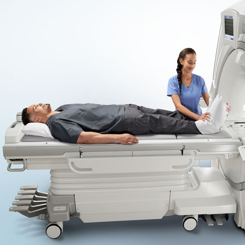
During MRI scanning, patients may have varying experiences influenced by factors such as their individual comfort levels, the duration of the scan, and the presence of claustrophobia. Here’s an overview of the patient experience during MRI scanning:
Preparation and Explanation: Before the MRI scan, patients are typically briefed by the healthcare team about the procedure. They are informed about what to expect during the scan, including the duration, the need to lie still, and the possibility of loud noises.
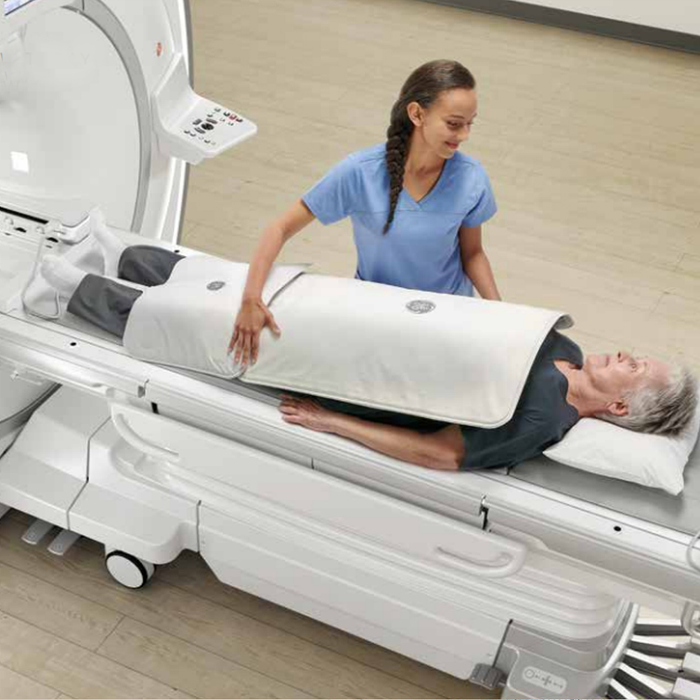
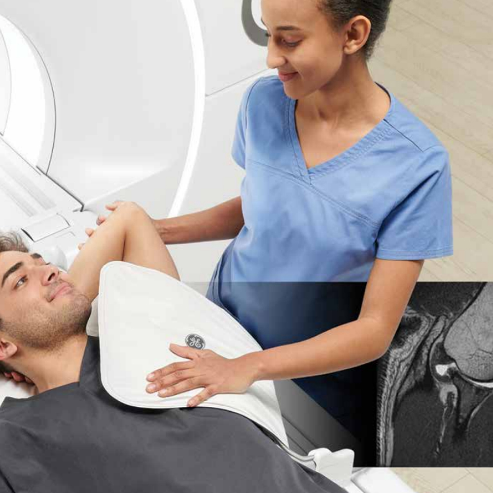
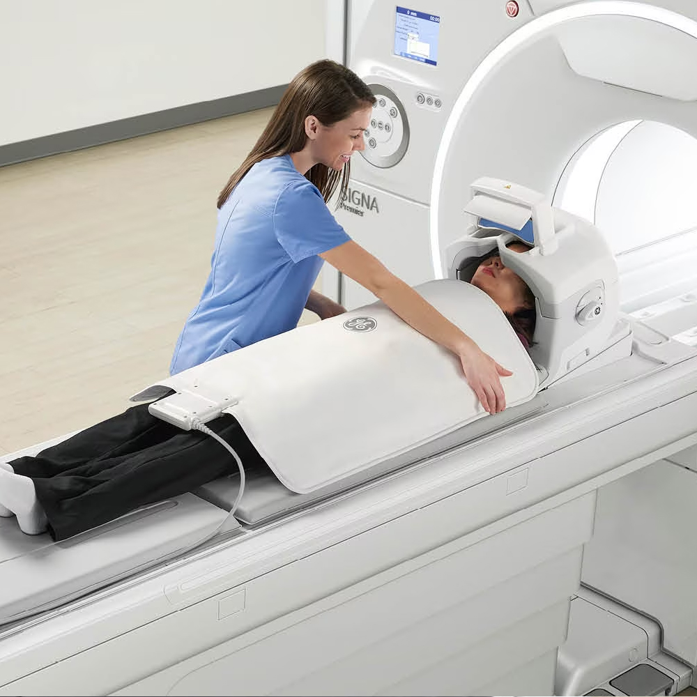
Positioning: Patients are positioned on a padded table that slides into the MRI machine. Depending on the area of the body being imaged, patients may need to lie flat on their back, stomach, or side.
Immobilization: Patients are often required to remain still during the MRI scan to avoid motion artifacts that could affect image quality. Some scans may require the use of immobilization devices or cushions to help patients maintain a comfortable position.
Noise: MRI scanners produce loud knocking or tapping noises during imaging, which can be unsettling for some patients. Earplugs or headphones are typically provided to help reduce the noise level and minimize discomfort.
Communication: Patients are usually in constant communication with the MRI technologist throughout the scan. They may be provided with a call button or intercom system to alert the technologist if they experience any discomfort or need assistance.
Claustrophobia: For patients with claustrophobia or anxiety, the confined space inside the MRI machine can be challenging. Open MRI machines or techniques such as sedation may be offered to alleviate anxiety and improve comfort.
Duration: MRI scans can range from a few minutes to over an hour, depending on the type of imaging being performed and the area of the body being examined. Patients are informed of the expected duration of the scan beforehand.
Comfort Measures: Healthcare providers may offer comfort measures such as blankets, pillows, or eye masks to enhance the patient’s comfort during the scan.
Post-Scan: After the scan is completed, patients are assisted in getting off the MRI table. They can resume their normal activities immediately unless sedation was used, in which case they may need some time to recover.
Overall, while MRI scanning can be intimidating for some patients, healthcare providers strive to ensure a positive and comfortable experience by addressing individual needs and concerns. Open communication, patient education, and the provision of comfort measures play crucial roles in enhancing the patient experience during MRI scanning.
3 Tesla Magnetic Resonance Imaging (3T MRI) refers to an advanced imaging technique that utilizes a magnetic field strength of 3 Tesla (T) to generate high-resolution images of the body’s internal structures. Here’s an overview of 3T MRI:
Enhanced Magnetic Field: Compared to traditional MRI machines with lower field strengths (e.g., 1.5T or lower), 3T MRI machines produce a stronger magnetic field, which results in increased signal-to-noise ratio and improved image quality.
Resolution and Detail: The higher magnetic field strength of 3T MRI allows for greater spatial resolution, enabling the visualization of smaller anatomical structures with enhanced detail. This makes 3T MRI particularly useful for imaging complex anatomical regions such as the brain, spine, joints, and blood vessels.
Clinical Applications: 3T MRI is utilized across various medical specialties for diagnostic imaging and research purposes. It is commonly used in neuroimaging for the detection and characterization of brain lesions, assessment of neurodegenerative diseases, and functional MRI (fMRI) studies. Additionally, it is employed in musculoskeletal imaging for evaluating joint disorders, soft tissue injuries, and musculoskeletal tumors.
Functional Imaging: The higher sensitivity of 3T MRI enables advanced functional imaging techniques such as diffusion tensor imaging (DTI), perfusion-weighted imaging (PWI), and magnetic resonance spectroscopy (MRS). These techniques provide valuable information about tissue microstructure, perfusion, and metabolite concentrations, aiding in the diagnosis and treatment planning of various medical conditions.
Clinical Considerations: While 3T MRI offers significant advantages in imaging quality and diagnostic capabilities, there are considerations to be mindful of, including potential safety concerns related to the higher magnetic field strength, patient comfort during longer scan times, and the need for specialized coils and protocols optimized for 3T imaging.
In summary, 3 Tesla Magnetic Resonance Imaging (3T MRI) is an advanced imaging modality that utilizes a higher magnetic field strength to achieve superior image resolution and diagnostic capabilities across a wide range of clinical applications. Its enhanced imaging quality makes it an invaluable tool for medical diagnosis, research, and treatment planning.
The characteristics and advantages of a 3 Tesla magnetic field in Magnetic Resonance Imaging (MRI) include:
Higher Signal-to-Noise Ratio (SNR): The 3 Tesla magnetic field strength provides a higher signal-to-noise ratio compared to lower field strengths. This results in clearer and more detailed images with improved contrast between tissues.
Increased Spatial Resolution: The higher magnetic field strength allows for increased spatial resolution, enabling the visualization of smaller anatomical structures with greater detail. This is particularly beneficial for imaging complex anatomical regions such as the brain, spine, and joints.
Enhanced Image Quality: 3 Tesla MRI produces images with higher image quality, providing better delineation of anatomical structures and improved diagnostic accuracy. This is advantageous for detecting subtle abnormalities and characterizing lesions.
Improved Functional Imaging: Higher field strength facilitates advanced functional imaging techniques such as functional MRI (fMRI), diffusion tensor imaging (DTI), and magnetic resonance spectroscopy (MRS). These techniques provide valuable insights into brain function, tissue microstructure, and metabolite concentrations.
Reduced Imaging Time: In some cases, 3 Tesla MRI can reduce imaging time compared to lower field strengths while maintaining image quality. This can improve patient comfort and throughput in clinical settings.
Enhanced Angiography: The higher magnetic field strength improves the visualization of blood vessels and enhances the quality of angiographic images. This is beneficial for assessing vascular anatomy and detecting abnormalities such as aneurysms and vascular malformations.
Research Applications: 3 Tesla MRI is widely used in research settings for studying various medical conditions and exploring novel imaging techniques. Its higher field strength enables researchers to investigate fine anatomical details and physiological processes with greater precision.
Clinical Versatility: While 3 Tesla MRI is particularly advantageous for neuroimaging and musculoskeletal imaging, it can also be applied across a wide range of clinical specialties, including oncology, cardiology, and abdominal imaging.
Overall, the 3 Tesla magnetic field in MRI offers several characteristics and advantages, including improved image quality, enhanced spatial resolution, and versatile clinical applications, making it a valuable tool for diagnostic imaging and research purposes.
The advantages of 3 Tesla Magnetic Resonance Imaging (MRI) compared to other magnetic field strengths, such as 1.5 Tesla (T) or lower, include:
Higher Signal-to-Noise Ratio (SNR): 3 Tesla MRI offers a higher signal-to-noise ratio compared to lower field strengths. This results in clearer and more detailed images with improved contrast between tissues, enhancing diagnostic accuracy.
Increased Spatial Resolution: The higher magnetic field strength of 3 Tesla MRI enables increased spatial resolution, allowing for better visualization of small anatomical structures and subtle lesions. This is particularly advantageous for imaging complex anatomical regions such as the brain, spine, and joints.
Enhanced Contrast: 3 Tesla MRI provides improved contrast between different tissue types, enhancing the ability to differentiate between normal and abnormal tissues. This is beneficial for detecting and characterizing lesions and abnormalities.
Improved Functional Imaging: Higher field strength facilitates advanced functional imaging techniques such as functional MRI (fMRI), diffusion tensor imaging (DTI), and magnetic resonance spectroscopy (MRS). These techniques provide valuable insights into brain function, tissue microstructure, and metabolite concentrations, aiding in diagnosis and treatment planning.
Reduced Imaging Time: In some cases, 3 Tesla MRI can reduce imaging time compared to lower field strengths while maintaining image quality. This can improve patient comfort and throughput in clinical settings.
Enhanced Angiography: The higher magnetic field strength of 3 Tesla MRI improves the visualization of blood vessels and enhances the quality of angiographic images. This is beneficial for assessing vascular anatomy and detecting abnormalities such as aneurysms and vascular malformations.
Clinical Versatility: While 3 Tesla MRI is particularly advantageous for neuroimaging and musculoskeletal imaging, it can also be applied across a wide range of clinical specialties, including oncology, cardiology, and abdominal imaging.
Overall, the higher magnetic field strength of 3 Tesla MRI offers several advantages over lower field strengths, including improved image quality, enhanced spatial resolution, and versatile clinical applications, making it a valuable tool for diagnostic imaging and research purposes.
The technology behind 3 Tesla Magnetic Resonance Imaging (MRI) involves several key components and advancements that contribute to its superior imaging capabilities. Here’s an overview of the technology of 3T MRI:
Magnet Design: 3 Tesla MRI machines feature high-strength superconducting magnets capable of generating a magnetic field strength of 3 Tesla. These magnets are designed with advanced materials and cooling systems to maintain their superconducting state, allowing for stable and uniform magnetic fields.
Gradient Coils: Gradient coils are used in 3T MRI machines to create spatial variations in the magnetic field, enabling spatial encoding of the MRI signals. These gradient coils are designed to produce high-amplitude and high-speed gradient pulses, allowing for rapid image acquisition and improved spatial resolution.
Radiofrequency (RF) Coils: RF coils are used to transmit and receive radiofrequency signals during MRI scanning. 3T MRI machines are equipped with specialized RF coils optimized for high-field imaging, which help improve signal sensitivity and image quality.
Parallel Imaging Techniques: Parallel imaging techniques such as sensitivity encoding (SENSE) and generalized autocalibrating partially parallel acquisitions (GRAPPA) are commonly used in 3T MRI to accelerate image acquisition. These techniques exploit the spatial information from multiple receiver coils to reduce scan times while maintaining image quality.
Advanced Pulse Sequences: 3T MRI utilizes advanced pulse sequences tailored to high-field imaging. These pulse sequences, including fast spin-echo, gradient echo, and echo-planar imaging, are optimized to take advantage of the higher signal-to-noise ratio and improved contrast available at 3 Tesla.
Parallel Transmit Technology: Some 3T MRI systems incorporate parallel transmit technology, also known as multi-transmit or multi-channel transmit, which utilizes multiple RF transmission channels to generate tailored RF pulses. This technology allows for improved uniformity of RF excitation and reduced artifacts in high-field imaging.
Image Reconstruction Algorithms: Sophisticated image reconstruction algorithms are employed in 3T MRI to process and reconstruct raw MRI data into high-resolution images. These algorithms account for factors such as gradient nonlinearity, magnetic field inhomogeneity, and RF coil sensitivity variations to ensure accurate image reconstruction.
Quality Assurance and Calibration: 3T MRI systems undergo rigorous quality assurance and calibration procedures to ensure optimal performance and image quality. Regular maintenance and calibration are essential to minimize system-related artifacts and maintain image consistency over time.
Overall, the technology of 3 Tesla MRI incorporates advanced magnet design, gradient coils, RF coils, parallel imaging techniques, pulse sequences, and image reconstruction algorithms to achieve superior imaging capabilities and diagnostic accuracy. These technological advancements contribute to the widespread adoption of 3T MRI in clinical practice and research.
A typical MRI system consists of several essential components that work together to acquire, process, and display magnetic resonance images. Here are the basic components of an MRI system:
Main Magnet: The main magnet is the core component of the MRI system, responsible for generating a strong magnetic field that aligns the hydrogen nuclei (protons) within the body. Most modern MRI systems use superconducting magnets, which require cryogenic cooling to maintain their superconducting state.
Gradient Coils: Gradient coils are additional coils positioned within the main magnet that produce small, spatially varying magnetic fields. These gradient fields are used to spatially encode the MRI signals, allowing for the determination of the location of signals within the body and the creation of detailed images.
Radiofrequency (RF) Coils: RF coils are used to transmit and receive radiofrequency signals during MRI scanning. These coils are positioned around the body part being imaged and are tuned to specific frequencies corresponding to the magnetic resonance of hydrogen nuclei. RF coils can be surface coils positioned close to the body or larger body coils that encircle the patient.
RF Transmitter: The RF transmitter is responsible for generating the RF pulses used to excite the hydrogen nuclei and produce magnetic resonance signals. These pulses are transmitted through the RF coils to the body part being imaged.
RF Receiver: The RF receiver detects the weak magnetic resonance signals emitted by the hydrogen nuclei in response to the RF pulses. These signals are then amplified, digitized, and processed to reconstruct images.
Gradient Amplifiers: Gradient amplifiers supply current to the gradient coils, generating the spatially varying magnetic fields necessary for spatial encoding of MRI signals. These amplifiers must deliver precise and rapid changes in current to produce the desired gradient fields.
Computer System: A computer system controls the operation of the MRI system, including the generation of RF pulses, control of gradient fields, acquisition of raw MRI data, and image reconstruction. Advanced software algorithms are used to process raw data and reconstruct high-resolution images.
Patient Table: The patient table is a movable platform on which the patient lies during MRI scanning. The table can be adjusted to position the patient correctly within the MRI system and move them in and out of the magnet bore.
Operator Console: The operator console is a workstation where the MRI technologist or radiologist controls the MRI system, monitors the scanning process, and interacts with the patient. The console provides user-friendly interfaces for selecting imaging parameters, viewing images, and adjusting scan protocols.
These are the basic components of an MRI system, each playing a crucial role in acquiring high-quality magnetic resonance images for diagnostic purposes. Additional accessories and safety features may also be included in modern MRI systems to ensure patient comfort and safety during scanning.
The operating principle and functioning of a 3 Tesla (3T) MRI scanner are based on the principles of magnetic resonance imaging (MRI) but with specific considerations for the higher magnetic field strength. Here’s an overview:
Main Magnet: The core component of a 3T MRI scanner is the main magnet, which generates a static magnetic field with a strength of 3 Tesla. This strong magnetic field aligns the nuclear spins of hydrogen atoms within the body.
Radiofrequency (RF) System: The RF system in a 3T MRI scanner includes RF coils for transmitting and receiving radiofrequency signals. These coils are tuned to the resonant frequency of hydrogen nuclei at 3T and are used to excite the spins and detect the resulting MR signals.
Gradient Coils: Gradient coils are used to spatially encode the MR signals, allowing for the creation of detailed images. In a 3T MRI scanner, high-performance gradient coils capable of producing strong and rapid gradient fields are utilized to achieve high-resolution imaging.
Pulse Sequences: Various pulse sequences, such as spin-echo, gradient echo, and echo-planar imaging, are employed in 3T MRI to manipulate the MR signals and generate different types of contrast and image contrasts.
Signal Reception and Processing: The MR signals detected by the RF coils are amplified, digitized, and processed by the MRI system. Advanced signal processing techniques are employed to reconstruct high-resolution images from the raw data.
Parallel Imaging: 3T MRI scanners often utilize parallel imaging techniques to accelerate image acquisition while maintaining image quality. These techniques exploit the spatial sensitivity of multiple receiver coils to reduce scan times.
Functional Imaging: 3T MRI scanners enable advanced functional imaging techniques, such as functional MRI (fMRI), diffusion tensor imaging (DTI), and magnetic resonance spectroscopy (MRS). These techniques provide valuable insights into brain function, tissue microstructure, and metabolite concentrations.
Safety Considerations: Due to the higher magnetic field strength of 3T MRI scanners, special considerations must be taken to ensure patient safety and minimize risks associated with the strong magnetic field, such as heating effects and peripheral nerve stimulation.
Overall, the operating principle and functioning of a 3T MRI scanner involve the utilization of a strong magnetic field, RF excitation, gradient encoding, and advanced imaging techniques to produce high-resolution images for diagnostic purposes.
The clinical applications and utilization of 3 Tesla (3T) Magnetic Resonance Imaging (MRI) scanners span across various medical specialties due to their high magnetic field strength and advanced imaging capabilities. Here are some key clinical applications and utilization of 3T MRI:
Neuroimaging:
- Brain Imaging: 3T MRI offers superior image resolution and contrast, making it ideal for detecting and characterizing brain lesions, tumors, vascular abnormalities, and neurodegenerative diseases such as Alzheimer’s disease.
- Functional MRI (fMRI): 3T MRI enables functional imaging of the brain, allowing for the mapping of brain activity during tasks and resting states, aiding in pre-surgical planning and research studies.
- Diffusion Tensor Imaging (DTI): 3T MRI is used for DTI to assess white matter integrity and connectivity in the brain, facilitating the diagnosis and monitoring of conditions such as stroke, multiple sclerosis, and traumatic brain injury.
Musculoskeletal Imaging:
- Joint Evaluation: 3T MRI provides detailed imaging of joints, cartilage, ligaments, and tendons, aiding in the diagnosis and management of conditions such as osteoarthritis, sports injuries, and ligament tears.
- Spine Imaging: 3T MRI offers high-resolution imaging of the spine, enabling the evaluation of spinal cord compression, disc herniation, spinal tumors, and degenerative spine diseases.
- Muscle Imaging: 3T MRI is used to assess muscle injuries, muscle atrophy, and muscle diseases such as muscular dystrophy.
Oncologic Imaging:
- Cancer Detection: 3T MRI is utilized for the detection, staging, and surveillance of various cancers, including breast cancer, prostate cancer, liver tumors, and head and neck cancers.
- Tumor Characterization: The high spatial resolution of 3T MRI aids in the characterization of tumors, distinguishing between benign and malignant lesions and assessing tumor size, location, and extent.
Cardiovascular Imaging:
- Cardiac MRI: 3T MRI is increasingly used for cardiac imaging, providing detailed assessment of cardiac anatomy, function, and myocardial perfusion. It is valuable for diagnosing and monitoring conditions such as myocardial infarction, cardiomyopathy, and congenital heart disease.
Abdominal and Pelvic Imaging:
- Liver Imaging: 3T MRI enables the assessment of liver morphology, vascularity, and liver lesions, including hepatocellular carcinoma and liver metastases.
- Pelvic Imaging: 3T MRI is used for imaging the pelvic organs, including the prostate, uterus, ovaries, and bladder, aiding in the diagnosis of gynecologic and urologic conditions.
Research Applications:
- 3T MRI is extensively utilized in research settings for studying neurological disorders, cognitive functions, musculoskeletal diseases, metabolic disorders, and cardiovascular conditions.
Overall, 3T MRI scanners have become indispensable tools in clinical practice, offering advanced imaging capabilities that enhance diagnostic accuracy, improve patient outcomes, and enable cutting-edge research in various medical specialties.
The role and applications of 3 Tesla (3T) Magnetic Resonance Imaging (MRI) in medical fields are extensive, owing to its high magnetic field strength and advanced imaging capabilities. Here are the key roles and applications of 3T MRI in various medical specialties:
Neurology:
- Brain Imaging: 3T MRI provides high-resolution imaging of the brain, enabling the detection and characterization of brain lesions, tumors, vascular abnormalities, and neurodegenerative diseases such as Alzheimer’s disease and multiple sclerosis.
- Functional MRI (fMRI): 3T MRI is used for functional brain imaging, allowing the mapping of brain activity during tasks and resting states. It is valuable for pre-surgical planning, research studies, and understanding brain function in neurological disorders.
Orthopedics and Musculoskeletal Imaging:
- Joint Evaluation: 3T MRI offers detailed imaging of joints, cartilage, ligaments, and tendons, aiding in the diagnosis and management of conditions such as osteoarthritis, sports injuries, ligament tears, and cartilage defects.
- Spine Imaging: 3T MRI provides high-resolution imaging of the spine, facilitating the evaluation of spinal cord compression, disc herniation, spinal tumors, and degenerative spine diseases.
- Muscle Imaging: 3T MRI is utilized for assessing muscle injuries, muscle atrophy, and muscle diseases such as muscular dystrophy.
Oncology:
- Cancer Detection and Staging: 3T MRI is used for the detection, staging, and surveillance of various cancers, including breast cancer, prostate cancer, liver tumors, and head and neck cancers.
- Tumor Characterization: 3T MRI aids in the characterization of tumors, distinguishing between benign and malignant lesions and assessing tumor size, location, and extent.
Cardiology:
- Cardiac MRI: 3T MRI is increasingly used for cardiac imaging, providing detailed assessment of cardiac anatomy, function, and myocardial perfusion. It is valuable for diagnosing and monitoring conditions such as myocardial infarction, cardiomyopathy, and congenital heart disease.
Abdominal and Pelvic Imaging:
- Liver and Abdominal Organs: 3T MRI enables detailed imaging of the liver and abdominal organs, aiding in the assessment of liver morphology, vascularity, liver lesions, and abdominal pathologies.
- Pelvic Imaging: 3T MRI is used for imaging pelvic organs such as the prostate, uterus, ovaries, and bladder, assisting in the diagnosis of gynecologic and urologic conditions.
Research and Advanced Imaging Techniques:
- 3T MRI is widely used in research settings for studying neurological disorders, cognitive functions, musculoskeletal diseases, metabolic disorders, and cardiovascular conditions.
- Advanced imaging techniques such as diffusion tensor imaging (DTI), functional MRI (fMRI), magnetic resonance spectroscopy (MRS), and perfusion-weighted imaging (PWI) are routinely performed on 3T MRI scanners for research and clinical purposes.
Overall, 3T MRI plays a crucial role in various medical fields by providing high-resolution imaging, advanced diagnostic capabilities, and valuable insights into a wide range of medical conditions.
The utilization of 3 Tesla (3T) Magnetic Resonance Imaging (MRI) in different body regions, including the brain, spine, joints, and other anatomical areas, is essential for diagnosing various medical conditions and assessing structural abnormalities. Here’s how 3T MRI is utilized in different body regions:
Brain Imaging:
- Structural Brain Imaging: 3T MRI provides high-resolution imaging of the brain, allowing for the detection and characterization of brain lesions, tumors, vascular abnormalities, and neurodegenerative diseases such as Alzheimer’s disease and multiple sclerosis.
- Functional MRI (fMRI): 3T MRI is used for functional brain imaging, enabling the mapping of brain activity during tasks and resting states. It is valuable for pre-surgical planning, research studies, and understanding brain function in neurological disorders.
Spine Imaging:
- Spinal Cord Compression: 3T MRI offers high-resolution imaging of the spine, facilitating the evaluation of spinal cord compression due to disc herniation, spinal stenosis, spinal tumors, and degenerative spine diseases.
- Disc Herniation: 3T MRI aids in the diagnosis and characterization of disc herniation, allowing for the assessment of disc protrusion, nerve root compression, and spinal canal narrowing.
- Degenerative Spine Diseases: 3T MRI is utilized for assessing degenerative spine diseases such as spondylosis, spondylolisthesis, and facet joint arthritis, aiding in treatment planning and patient management.
Joint Imaging:
- Knee Imaging: 3T MRI provides detailed imaging of the knee joint, allowing for the evaluation of meniscal tears, ligament injuries (e.g., ACL tears), cartilage defects, and osteoarthritis.
- Shoulder Imaging: 3T MRI aids in the assessment of shoulder pathology, including rotator cuff tears, labral tears, glenohumeral instability, and shoulder impingement syndrome.
- Hip Imaging: 3T MRI enables the diagnosis of hip pathology such as labral tears, femoroacetabular impingement (FAI), hip dysplasia, and avascular necrosis (AVN).
Musculoskeletal Imaging:
- Muscle and Soft Tissue Evaluation: 3T MRI is used for assessing muscle injuries, muscle atrophy, soft tissue tumors, and inflammatory conditions such as myositis.
- Bone and Joint Disorders: 3T MRI aids in the diagnosis and characterization of bone and joint disorders, including osteoarthritis, rheumatoid arthritis, bone tumors, and stress fractures.
Other Body Regions:
- Abdominal Imaging: 3T MRI enables detailed imaging of abdominal organs such as the liver, pancreas, kidneys, and spleen, aiding in the diagnosis of liver lesions, pancreatic tumors, renal masses, and abdominal pathologies.
- Pelvic Imaging: 3T MRI is utilized for imaging pelvic organs such as the prostate, uterus, ovaries, and bladder, assisting in the diagnosis of gynecologic and urologic conditions.
Overall, 3T MRI is extensively utilized in various body regions for diagnosing medical conditions, assessing structural abnormalities, and guiding treatment decisions in clinical practice.
The advantages of 3 Tesla (3T) Magnetic Resonance Imaging (MRI) compared to lower magnetic field strengths, such as 1.5 Tesla (1.5T) or lower, include:
Improved Image Quality: The higher magnetic field strength of 3T MRI results in improved signal-to-noise ratio (SNR), leading to higher image quality with better contrast resolution and spatial resolution. This allows for enhanced visualization of anatomical structures and pathological abnormalities.
Enhanced Spatial Resolution: 3T MRI provides greater spatial resolution compared to lower field strengths, enabling the visualization of smaller anatomical details and subtle lesions. This is particularly advantageous for imaging complex anatomical regions and for detecting small abnormalities.
Better Tissue Contrast: The higher SNR and improved contrast resolution of 3T MRI contribute to better tissue characterization and differentiation between normal and abnormal tissues. This is beneficial for detecting and characterizing lesions, tumors, and other pathological changes.
Faster Imaging: In some cases, 3T MRI allows for faster imaging compared to lower field strengths, while still maintaining high image quality. This can lead to shorter scan times, increased patient throughput, and improved patient comfort during imaging procedures.
Advanced Functional Imaging: 3T MRI facilitates advanced functional imaging techniques, such as functional MRI (fMRI), diffusion tensor imaging (DTI), and magnetic resonance spectroscopy (MRS). These techniques provide valuable insights into brain function, tissue microstructure, and metabolic activity.
Improved Angiography: The higher magnetic field strength of 3T MRI enhances the visualization of blood vessels and improves the quality of angiographic images. This is beneficial for assessing vascular anatomy, detecting vascular lesions, and evaluating blood flow dynamics.
Versatility in Applications: 3T MRI is versatile and can be applied across a wide range of clinical specialties, including neurology, orthopedics, oncology, cardiology, and abdominal imaging. Its high image quality and advanced imaging capabilities make it valuable for diagnosing various medical conditions and guiding treatment decisions.
Research Capabilities: 3T MRI is widely used in research settings for studying neurological disorders, musculoskeletal diseases, oncology, and other medical conditions. Its higher magnetic field strength allows for advanced research applications and the development of novel imaging techniques.
Overall, the advantages of 3T MRI, including improved image quality, enhanced spatial resolution, better tissue contrast, faster imaging, advanced functional imaging capabilities, versatility in applications, and research capabilities, make it a valuable tool in clinical practice and research.
One of the significant advantages of 3 Tesla (3T) Magnetic Resonance Imaging (MRI) is its capability to provide high resolution and contrast in imaging studies. Here’s how 3T MRI achieves high resolution and contrast:
Higher Signal-to-Noise Ratio (SNR): The higher magnetic field strength of 3T MRI results in an increased signal-to-noise ratio compared to lower field strengths, such as 1.5T or lower. This higher SNR allows for better image quality with improved spatial resolution and contrast.
Improved Spatial Resolution: The higher magnetic field strength of 3T MRI enables the use of smaller voxels (volume elements) during image acquisition, leading to higher spatial resolution. This allows for the visualization of smaller anatomical structures and subtle pathological changes with greater detail.
Enhanced Contrast Resolution: 3T MRI provides better contrast resolution, allowing for improved differentiation between different types of tissues. This enhanced contrast resolution is particularly beneficial for distinguishing between normal and abnormal tissues, detecting lesions, and characterizing pathological changes.
Advanced Pulse Sequences: 3T MRI utilizes advanced pulse sequences optimized for high-field imaging, which contribute to improved image resolution and contrast. These pulse sequences, such as fast spin-echo, gradient echo, and echo-planar imaging, are tailored to take advantage of the higher SNR and contrast available at 3T.
Parallel Imaging Techniques: Parallel imaging techniques, such as sensitivity encoding (SENSE) and generalized autocalibrating partially parallel acquisitions (GRAPPA), are commonly used in 3T MRI to accelerate image acquisition while maintaining image quality. These techniques exploit the spatial information from multiple receiver coils to improve image resolution and contrast.
Higher Field Strength Benefits: The inherent properties of higher magnetic field strength, such as increased T1 and T2 relaxation times, contribute to improved image contrast in 3T MRI. This results in better differentiation between tissues with varying relaxation times, enhancing contrast in the final images.
Overall, the combination of higher SNR, improved spatial resolution, advanced pulse sequences, parallel imaging techniques, and the benefits of higher magnetic field strength enables 3T MRI to achieve high resolution and contrast, making it a valuable tool for diagnostic imaging across various medical specialties.
One of the advantages of 3 Tesla (3T) Magnetic Resonance Imaging (MRI) is its capability to offer faster scan times and reduced waiting periods for patients undergoing imaging studies. Here’s how 3T MRI achieves this:
Improved Signal-to-Noise Ratio (SNR): The higher magnetic field strength of 3T MRI results in an increased signal-to-noise ratio compared to lower field strengths, such as 1.5T or lower. This higher SNR allows for faster image acquisition with shorter scan times while maintaining image quality.
Advanced Pulse Sequences: 3T MRI utilizes advanced pulse sequences optimized for high-field imaging, which contribute to faster image acquisition. These pulse sequences, such as echo-planar imaging (EPI) and fast spin-echo, are designed to acquire multiple image slices in a single scan, reducing the overall scan time.
Parallel Imaging Techniques: Parallel imaging techniques, such as sensitivity encoding (SENSE) and generalized autocalibrating partially parallel acquisitions (GRAPPA), are commonly used in 3T MRI to accelerate image acquisition. These techniques exploit the spatial information from multiple receiver coils to reduce scan times while maintaining image quality.
Higher Gradient Performance: 3T MRI scanners often feature high-performance gradient systems capable of producing strong and rapid gradient fields. These gradient coils allow for faster spatial encoding of the MRI signals, enabling shorter echo times and faster image acquisition.
Optimized Imaging Protocols: Radiologists and technologists can optimize imaging protocols for 3T MRI to achieve faster scan times while meeting diagnostic requirements. This includes selecting appropriate pulse sequences, imaging parameters, and coil configurations to balance scan speed with image quality.
Efficient Workflow and System Design: 3T MRI systems are designed with features that enhance workflow efficiency, such as user-friendly interfaces, automated scan protocols, and streamlined patient positioning and setup. These features help minimize downtime between scans and reduce waiting periods for patients.
Increased Patient Throughput: The combination of faster scan times and efficient workflow in 3T MRI enables increased patient throughput, allowing more patients to be imaged within a given time frame. This helps reduce waiting periods for scheduling appointments and improves overall patient satisfaction.
Overall, the combination of improved SNR, advanced pulse sequences, parallel imaging techniques, optimized imaging protocols, efficient workflow, and increased patient throughput in 3T MRI contributes to faster scan times and reduced waiting periods for patients undergoing MRI examinations.
Information obtained with 3 Tesla (3T) Magnetic Resonance Imaging (MRI) is extensive and provides detailed insights into various anatomical structures and pathological conditions. Here are some types of information that can be obtained with 3T MRI:
Anatomical Detail:
- High-resolution imaging: 3T MRI offers superior spatial resolution, allowing for detailed visualization of anatomical structures such as the brain, spine, joints, and organs.
- Fine tissue differentiation: With improved contrast resolution, 3T MRI can distinguish between different types of tissues, aiding in the characterization of normal and abnormal structures.
Pathological Conditions:
- Lesion detection: 3T MRI is sensitive to detecting lesions, tumors, cysts, and other abnormalities in various body regions, including the brain, spine, musculoskeletal system, and organs.
- Tumor characterization: 3T MRI provides detailed information about the size, location, morphology, and vascularity of tumors, aiding in their characterization and staging.
Functional Information:
- Functional MRI (fMRI): 3T MRI enables functional imaging of the brain, allowing for the mapping of brain activity during tasks and resting states. This information is valuable for understanding brain function and identifying regions involved in specific cognitive processes.
- Diffusion Tensor Imaging (DTI): 3T MRI is used for DTI to assess white matter integrity and connectivity in the brain, providing insights into neurological disorders, such as stroke, multiple sclerosis, and traumatic brain injury.
Vascular Imaging:
- Angiography: 3T MRI provides high-resolution angiographic images of blood vessels, enabling the evaluation of vascular anatomy, detecting stenosis, aneurysms, and vascular malformations, and assessing blood flow dynamics.
Musculoskeletal Assessment:
- Joint evaluation: 3T MRI offers detailed imaging of joints, ligaments, tendons, and cartilage, facilitating the diagnosis and management of orthopedic conditions such as osteoarthritis, sports injuries, and ligament tears.
- Muscle assessment: 3T MRI is used to evaluate muscle injuries, muscle atrophy, inflammatory myopathies, and neuromuscular disorders.
Cardiovascular Evaluation:
- Cardiac MRI: 3T MRI enables comprehensive assessment of cardiac anatomy, function, myocardial perfusion, and tissue characterization, aiding in the diagnosis and management of cardiovascular diseases.
Abdominal and Pelvic Imaging:
- Abdominal organs: 3T MRI provides detailed imaging of abdominal organs such as the liver, pancreas, kidneys, spleen, and gastrointestinal tract, aiding in the diagnosis of liver lesions, pancreatic tumors, renal masses, and abdominal pathologies.
- Pelvic organs: 3T MRI is utilized for imaging pelvic organs such as the prostate, uterus, ovaries, and bladder, assisting in the diagnosis of gynecologic and urologic conditions.
Overall, 3T MRI offers a comprehensive range of information, including anatomical detail, pathological conditions, functional information, vascular imaging, musculoskeletal assessment, cardiovascular evaluation, and abdominal and pelvic imaging, making it a versatile tool for diagnostic imaging across various medical specialties.
The ability of 3 Tesla (3T) Magnetic Resonance Imaging (MRI) for anatomical and pathological assessment is unparalleled due to its high spatial resolution, excellent tissue contrast, and advanced imaging capabilities. Here’s how 3T MRI facilitates anatomical and pathological assessment:
Anatomical Assessment:
- High Spatial Resolution: 3T MRI offers superior spatial resolution compared to lower field strengths, allowing for detailed visualization of anatomical structures with excellent clarity and precision.
- Multiplanar Imaging: 3T MRI enables imaging in multiple planes (sagittal, coronal, and axial), providing comprehensive views of anatomical structures from different perspectives.
- Soft Tissue Differentiation: With its excellent tissue contrast, 3T MRI can distinguish between different types of soft tissues, enhancing the delineation of anatomical boundaries and structures.
Pathological Assessment:
- Lesion Detection: 3T MRI is highly sensitive to detecting pathological lesions, including tumors, cysts, abscesses, and inflammatory lesions, in various body regions such as the brain, spine, joints, and organs.
- Tumor Characterization: 3T MRI provides detailed information about the morphology, size, location, vascularity, and composition of tumors, aiding in their characterization, staging, and treatment planning.
- Assessment of Disease Progression: 3T MRI enables longitudinal assessment of disease progression by accurately detecting and monitoring pathological changes over time, allowing for timely intervention and management.
Functional Correlation:
- Functional MRI (fMRI): In addition to anatomical assessment, 3T MRI facilitates functional assessment of the brain through fMRI, which maps brain activity in response to specific stimuli or tasks. This functional correlation provides valuable insights into brain function and connectivity.
- Diffusion Tensor Imaging (DTI): 3T MRI is utilized for DTI to assess white matter integrity and connectivity in the brain, correlating structural abnormalities with functional deficits in neurological disorders.
Quantitative Assessment:
- Morphometric Analysis: 3T MRI allows for quantitative assessment of anatomical structures through morphometric analysis, measuring parameters such as volumes, areas, and distances, which are valuable for research and clinical purposes.
- Quantitative Diffusion Imaging: With its advanced diffusion imaging techniques, such as diffusion-weighted imaging (DWI) and diffusion tensor imaging (DTI), 3T MRI enables quantitative assessment of tissue microstructure and integrity, aiding in the characterization of pathological changes.
Overall, 3T MRI provides unparalleled capabilities for anatomical and pathological assessment, offering high spatial resolution, excellent tissue contrast, functional correlation, and quantitative analysis, making it a valuable tool for diagnostic imaging and research in various medical specialties.
The ability of 3 Tesla (3T) Magnetic Resonance Imaging (MRI) to provide functional information is a key advantage of this imaging modality. Here’s how 3T MRI facilitates the assessment of functional information:
Functional MRI (fMRI):
- Brain Function Mapping: 3T MRI is widely used for functional MRI (fMRI) studies to map brain activity in response to various stimuli or tasks. By detecting changes in blood oxygenation levels associated with neuronal activity (BOLD effect), fMRI helps identify brain regions involved in specific cognitive functions such as language, motor control, memory, and sensory processing.
- Resting-State fMRI: 3T MRI can also assess brain function during resting states, providing insights into intrinsic brain networks and connectivity patterns. Resting-state fMRI is valuable for studying brain disorders and understanding the brain’s functional organization.
Diffusion Tensor Imaging (DTI):
- White Matter Tractography: 3T MRI facilitates diffusion tensor imaging (DTI), a technique that measures the diffusion of water molecules in brain tissue. DTI enables the visualization and mapping of white matter tracts in the brain, providing information about structural connectivity and integrity. DTI is used to study conditions affecting white matter, such as traumatic brain injury, stroke, and neurodegenerative diseases.
Magnetic Resonance Spectroscopy (MRS):
- Metabolic Imaging: 3T MRI offers magnetic resonance spectroscopy (MRS), a technique that measures chemical metabolites in tissues. MRS provides information about tissue metabolism and biochemistry, aiding in the assessment of various neurological and oncological conditions. For example, MRS can detect alterations in brain metabolites associated with tumors, ischemia, and neurodegenerative disorders.
Arterial Spin Labeling (ASL):
- Cerebral Blood Flow Mapping: 3T MRI can perform arterial spin labeling (ASL), a non-invasive technique for measuring cerebral blood flow (CBF) in the brain. ASL provides quantitative information about regional perfusion, which is valuable for studying brain disorders such as stroke, dementia, and cerebrovascular diseases.
Functional Connectivity MRI (fcMRI):
- Brain Network Analysis: 3T MRI enables functional connectivity MRI (fcMRI), a technique that assesses synchronized activity between different brain regions, revealing functional brain networks. fcMRI helps identify alterations in brain connectivity associated with neurological and psychiatric disorders, providing insights into disease mechanisms and treatment effects.
Overall, 3T MRI offers advanced functional imaging capabilities, including fMRI, DTI, MRS, ASL, and fcMRI, which provide valuable information about brain function, tissue metabolism, perfusion, and connectivity. These functional imaging techniques are essential for understanding normal brain function, identifying abnormalities, and guiding treatment strategies in various neurological and psychiatric conditions.
3 Tesla (3T) Magnetic Resonance Imaging (MRI) is generally considered safe when performed under appropriate conditions and guidelines. However, there are certain safety considerations to be aware of:
Magnetic Field Hazards:
- Strong magnetic fields: 3T MRI scanners produce a strong magnetic field, which can pose hazards for individuals with certain medical devices or metallic implants. It is essential to screen patients for contraindications before undergoing 3T MRI to ensure safety.
- Projectile hazards: Ferromagnetic objects can become projectiles in the magnetic field of an MRI scanner. Strict safety protocols are in place to prevent accidents, including screening individuals for metallic objects and ensuring the MRI suite is free of ferromagnetic materials.
Radiofrequency (RF) Heating:
- RF heating: The RF pulses used in MRI can cause heating of tissues, particularly in regions with high conductivity (e.g., tissues with high water content). Safety guidelines are in place to limit RF exposure and prevent excessive heating during MRI scans.
- Monitoring: Continuous monitoring of patient temperature and physiological parameters during MRI scans helps ensure patient safety and detect any signs of RF heating.
Contrast Agents:
- Gadolinium-based contrast agents: While gadolinium-based contrast agents are commonly used in MRI to enhance image quality, they carry a risk of adverse reactions, including allergic reactions and nephrogenic systemic fibrosis (NSF) in patients with impaired renal function. Precautions are taken to minimize risks associated with contrast administration, including screening for contraindications and monitoring patients for adverse reactions.
Acoustic Noise:
- Acoustic noise: MRI scans produce loud noises due to the switching of gradient coils, which can be uncomfortable for patients and may require ear protection. Safety protocols include providing earplugs or headphones to reduce noise exposure during MRI scans.
Patient Monitoring:
- Patient monitoring: Continuous monitoring of patients’ vital signs and physiological parameters during MRI scans is essential to ensure their safety and well-being. Qualified healthcare personnel should be present to monitor patients throughout the procedure and respond to any emergent situations.
Pregnancy and Pediatrics:
- Safety considerations for pregnant women and pediatric patients: Special precautions are taken for pregnant women and pediatric patients undergoing MRI to minimize potential risks. MRI is generally considered safe during pregnancy when medically necessary, but specific safety guidelines and protocols are followed to ensure fetal safety.
Overall, 3T MRI is considered safe when performed by trained healthcare professionals following established safety guidelines and protocols. Precautions are taken to minimize risks associated with the magnetic field, RF heating, contrast agents, acoustic noise, and patient monitoring, ensuring the safety and well-being of patients undergoing MRI scans.
Several precautions are taken to ensure patient safety during scanning in a 3 Tesla (3T) Magnetic Resonance Imaging (MRI) environment. These precautions are essential to minimize risks associated with the magnetic field, radiofrequency (RF) energy, contrast agents, and other potential hazards. Here are some key precautions:
Screening for Contraindications:
- Patients are thoroughly screened for contraindications to MRI, including metallic implants, medical devices, pacemakers, cochlear implants, aneurysm clips, and other ferromagnetic objects that may pose hazards in the magnetic field.
Patient Education:
- Patients are provided with detailed information about the MRI procedure, including safety guidelines, potential risks, and instructions to follow during the scan (e.g., remaining still, avoiding movement).
Metal Detection:
- Prior to entering the MRI suite, patients are screened for metallic objects using metal detectors to ensure they do not have any ferromagnetic materials that could pose projectile hazards in the magnetic field.
Patient Monitoring:
- Continuous monitoring of patients’ vital signs (e.g., heart rate, blood pressure, oxygen saturation) and physiological parameters is performed throughout the MRI scan to ensure their safety and well-being.
RF Heating Monitoring:
- RF heating levels are monitored during the MRI scan to prevent excessive tissue heating, particularly in regions with high conductivity. Temperature monitoring devices may be used to detect any signs of RF heating in vulnerable patients.
Contrast Agent Safety:
- If contrast agents (e.g., gadolinium-based contrast agents) are administered for enhanced imaging, patients are screened for contraindications (e.g., history of allergic reactions, impaired renal function) to minimize risks. Appropriate dosage and administration protocols are followed to ensure patient safety.
Ear Protection:
- Patients are provided with earplugs or headphones to protect against the loud acoustic noise generated by the MRI scanner during image acquisition.
Patient Comfort:
- Measures are taken to ensure patient comfort during the MRI scan, including providing blankets, pillows, and communication devices to alleviate anxiety and promote relaxation.
Emergency Preparedness:
- Healthcare personnel are trained in emergency response procedures and equipped to handle any medical emergencies that may arise during the MRI scan, including adverse reactions to contrast agents or unexpected physiological changes in patients.
Patient Assistance:
- Patients are reassured and supported throughout the MRI scan by trained healthcare professionals who are available to address any concerns or questions they may have during the procedure.
By adhering to these precautions and safety measures, healthcare providers can ensure the safe and effective performance of MRI scans for patients undergoing imaging studies in a 3T MRI environment.
The evolution of 3 Tesla (3T) Magnetic Resonance Imaging (MRI) technology has led to a changing role in clinical applications, with advancements in imaging capabilities, diagnostic accuracy, and therapeutic guidance. Here’s how the role of 3T MRI has evolved and continues to change in clinical applications:
Improved Image Quality:
- Evolution: Advances in hardware and software technology have enhanced the image quality of 3T MRI, providing higher spatial resolution, improved tissue contrast, and better anatomical detail compared to lower field strengths.
- Changing Role: With improved image quality, 3T MRI has become increasingly valuable for diagnosing subtle abnormalities, characterizing lesions, and guiding treatment decisions in various medical specialties.
Expanded Clinical Applications:
- Evolution: Initially used primarily in neuroimaging applications, 3T MRI has expanded its clinical applications to include musculoskeletal imaging, cardiovascular imaging, abdominal imaging, and oncological imaging, among others.
- Changing Role: The versatility of 3T MRI across multiple medical specialties has led to its broader adoption in clinical practice, offering a comprehensive imaging solution for diagnosing and managing a wide range of medical conditions.
Functional Imaging Capabilities:
- Evolution: 3T MRI technology enables advanced functional imaging techniques such as functional MRI (fMRI), diffusion tensor imaging (DTI), magnetic resonance spectroscopy (MRS), and arterial spin labeling (ASL), providing valuable insights into brain function, tissue microstructure, and metabolic activity.
- Changing Role: Functional imaging with 3T MRI has become essential for understanding neurological disorders, mapping brain function, evaluating treatment response, and advancing research in neuroscience and neuropsychology.
Therapeutic Guidance:
- Evolution: 3T MRI is increasingly used for therapeutic guidance in interventional procedures, surgical planning, and monitoring treatment response. Real-time imaging capabilities enable precise localization of targets, accurate needle placement, and intraoperative visualization of anatomical structures.
- Changing Role: 3T MRI plays a crucial role in minimally invasive procedures, such as neurosurgery, orthopedic surgery, oncological interventions, and cardiovascular interventions, where precise anatomical guidance is essential for optimal patient outcomes.
Research and Innovation:
- Evolution: 3T MRI continues to drive research and innovation in medical imaging, fueling developments in image acquisition techniques, image analysis algorithms, and novel imaging biomarkers.
- Changing Role: As a research tool, 3T MRI enables investigators to explore new imaging modalities, investigate disease mechanisms, validate imaging biomarkers, and develop personalized diagnostic and therapeutic strategies for precision medicine.
Clinical Integration and Workflow Optimization:
- Evolution: Integration of 3T MRI into clinical workflows has evolved with advancements in scanner design, workflow optimization, and streamlined image acquisition protocols.
- Changing Role: Efforts to improve patient throughput, reduce scan times, and enhance workflow efficiency have made 3T MRI more accessible and practical in clinical settings, leading to increased utilization and improved patient care.
Overall, the evolution of 3T MRI technology has resulted in a changing role in clinical applications, expanding its scope beyond traditional neuroimaging to encompass a wide range of medical specialties, functional imaging modalities, therapeutic guidance, research innovation, and workflow optimization, thereby contributing to improved patient care and outcomes.
Future directions and potential improvements in 3 Tesla (3T) Magnetic Resonance Imaging (MRI) technology are driven by ongoing research, technological advancements, and clinical needs. Here are some areas of focus and potential improvements in the future of 3T MRI:
Higher Field Strengths:
- Future advancements may involve the development of even higher field strengths beyond 3T, such as 7T or higher. Higher field strengths offer the potential for increased signal-to-noise ratio (SNR), improved spatial resolution, and enhanced sensitivity for detecting subtle anatomical and pathological features.
Advanced Imaging Sequences:
- Continued research and development of advanced imaging sequences tailored for specific clinical applications are expected. This includes optimizing pulse sequences for improved tissue contrast, reduced artifacts, faster scan times, and enhanced functional and metabolic imaging capabilities.
Functional and Molecular Imaging:
- Future advancements in functional and molecular imaging techniques using 3T MRI may enable more detailed characterization of physiological processes, cellular metabolism, and molecular pathways. This includes advancements in techniques such as functional MRI (fMRI), diffusion tensor imaging (DTI), magnetic resonance spectroscopy (MRS), and molecular imaging probes.
Artificial Intelligence (AI) Integration:
- Integration of artificial intelligence (AI) and machine learning algorithms into 3T MRI data analysis may lead to automated image processing, quantitative analysis, and diagnostic decision support. AI-based tools could improve workflow efficiency, enhance diagnostic accuracy, and facilitate personalized treatment planning.
Quantitative Imaging Biomarkers:
- Development of quantitative imaging biomarkers derived from 3T MRI data may provide valuable information for disease diagnosis, prognosis, and treatment response assessment. Quantitative biomarkers related to tissue microstructure, perfusion, diffusion, and metabolism could improve clinical decision-making and patient management.
Multimodal Imaging Integration:
- Integration of 3T MRI with other imaging modalities, such as positron emission tomography (PET), computed tomography (CT), and ultrasound, may enable comprehensive multimodal imaging assessments. Combining complementary imaging modalities could provide synergistic diagnostic information and enhance the understanding of complex diseases.
Patient-Centered Design:
- Future improvements in 3T MRI scanner design may focus on enhancing patient comfort, reducing acoustic noise levels, and minimizing claustrophobia during imaging procedures. Open-bore and wide-bore MRI designs, as well as immersive audiovisual experiences, could improve the patient experience and compliance.
Quantitative Susceptibility Mapping (QSM):
- Further development and clinical implementation of quantitative susceptibility mapping (QSM) techniques at 3T MRI may improve the assessment of tissue iron content, neurodegenerative diseases, and cerebrovascular disorders. QSM provides quantitative information about tissue magnetic susceptibility, offering insights into brain physiology and pathology.
Intraoperative MRI (iMRI):
- Integration of 3T MRI into intraoperative settings may enable real-time imaging guidance during surgical procedures. iMRI systems with high-field strength could provide surgeons with updated anatomical information, facilitate tumor resection, and improve surgical outcomes in neurosurgery and other specialties.
Remote Monitoring and Telemetry:
- Advancements in remote monitoring and telemetry technologies may allow for real-time monitoring of patients undergoing 3T MRI scans, including physiological parameters, patient motion, and device status. Remote monitoring systems could enhance patient safety and enable remote assistance from healthcare providers during imaging procedures.
Overall, the future of 3T MRI holds promise for continued advancements in imaging technology, clinical applications, and patient care, driven by ongoing research, interdisciplinary collaboration, and technological innovation.
When undergoing Magnetic Resonance Imaging (MRI) scanning, patients have certain rights and considerations to ensure their safety, comfort, and informed participation in the procedure. Here are some important considerations and patient rights regarding MRI scanning:
Informed Consent:
- Patients have the right to receive clear and comprehensive information about the MRI procedure, including its purpose, risks, benefits, alternatives, and potential complications. Informed consent ensures that patients understand the procedure and can make voluntary decisions about their healthcare.
Safety Screening:
- Patients should undergo thorough safety screening before the MRI scan to identify any contraindications, such as metallic implants, medical devices, or other conditions that may pose risks in the magnetic field. Safety screening ensures patient safety and prevents potential hazards during the procedure.
Privacy and Confidentiality:
- Patients have the right to privacy and confidentiality regarding their medical information and imaging results. Healthcare providers should adhere to strict privacy protocols to protect patient confidentiality and ensure that only authorized individuals have access to the patient’s MRI data.
Comfort and Support:
- Patients have the right to receive adequate support and accommodations to ensure their comfort during the MRI scan. This may include providing pillows, blankets, earplugs, or headphones to minimize discomfort from the loud noise generated by the MRI scanner. Patients with claustrophobia or anxiety may benefit from additional support and reassurance from healthcare providers.
Communication and Education:
- Patients should receive clear and understandable explanations about the MRI procedure, including what to expect during the scan, how to prepare for it, and any instructions to follow before, during, and after the procedure. Effective communication helps alleviate anxiety and ensures that patients feel informed and empowered throughout the process.
Patient Advocacy:
- Patients have the right to advocate for their own healthcare needs and preferences during the MRI scan. This includes expressing any concerns, preferences, or requests related to the procedure, such as the need for breaks, adjustments in positioning, or accommodations for physical limitations.
Emergency Preparedness:
- Healthcare providers should be prepared to respond promptly to any medical emergencies or adverse reactions that may occur during the MRI scan. This includes having emergency protocols in place, trained personnel available on-site, and access to emergency medical equipment and medications.
Respect for Dignity and Autonomy:
- Patients have the right to be treated with dignity, respect, and autonomy throughout the MRI scanning process. Healthcare providers should recognize and honor patients’ autonomy, cultural beliefs, preferences, and personal boundaries, ensuring that they feel respected and valued as individuals.
Feedback and Follow-Up:
- Patients should have the opportunity to provide feedback about their MRI experience and raise any concerns or questions they may have. Healthcare providers should address patient feedback promptly and provide appropriate follow-up care or referrals as needed.
Patient Rights Advocacy:
- Patients have the right to advocate for their rights and interests during the MRI scanning process. This may involve seeking support from patient advocacy organizations, legal resources, or healthcare professionals to ensure that their rights are upheld and respected.
Overall, patients undergoing MRI scanning have the right to receive safe, respectful, and patient-centered care that prioritizes their well-being, comfort, and informed participation in the procedure. Healthcare providers should adhere to ethical principles, professional standards, and legal regulations to ensure that patients’ rights and considerations are respected throughout the MRI scanning process.
Privacy and data protection are critical considerations in Magnetic Resonance Imaging (MRI) scanning to safeguard patients’ sensitive medical information and imaging data. Here are some key aspects related to privacy and data protection in MRI scanning:
Patient Confidentiality:
- Patient confidentiality is paramount in MRI scanning. Healthcare providers must ensure that patients’ personal and medical information remains confidential and is only accessed by authorized personnel involved in the patient’s care.
Data Encryption:
- MRI imaging data should be encrypted during transmission and storage to protect it from unauthorized access or interception. Encryption helps prevent data breaches and ensures that patient information remains secure.
Access Controls:
- Access to MRI imaging data should be restricted to authorized healthcare professionals who have a legitimate need to view or use the data for patient care purposes. Access controls, such as user authentication and role-based permissions, help prevent unauthorized access to patient data.
Secure Storage:
- MRI imaging data should be stored securely in compliance with data protection regulations and industry best practices. Secure storage systems, including encrypted servers and cloud storage solutions, help protect patient data from unauthorized access, loss, or theft.
Data Retention Policies:
- Healthcare providers should establish data retention policies for MRI imaging data, specifying the duration for which data will be retained and the procedures for securely deleting or archiving data when it is no longer needed for patient care or legal purposes.
Patient Consent:
- Patients should provide informed consent for the collection, storage, and use of their MRI imaging data for diagnostic and treatment purposes. Healthcare providers should explain the purposes and potential risks associated with MRI imaging and obtain patients’ consent before conducting the procedure.
Compliance with Regulations:
- Healthcare providers must comply with data protection regulations, such as the Health Insurance Portability and Accountability Act (HIPAA) in the United States, the General Data Protection Regulation (GDPR) in the European Union, and other relevant laws and regulations governing patient privacy and data security.
Data Breach Response Plan:
- Healthcare providers should have a data breach response plan in place to address potential security incidents involving MRI imaging data. The plan should outline procedures for detecting, reporting, and responding to data breaches, as well as notifying affected individuals and regulatory authorities as required by law.
Staff Training:
- Healthcare staff involved in MRI scanning should receive training on privacy and data protection policies and procedures. Training helps ensure that staff understand their responsibilities for safeguarding patient data and are aware of best practices for maintaining patient confidentiality.
Audit Trails and Monitoring:
- Healthcare providers should implement audit trails and monitoring mechanisms to track access to MRI imaging data and detect any unauthorized or suspicious activities. Audit trails help identify potential security breaches and ensure accountability for data access and usage.
By addressing these aspects of privacy and data protection, healthcare providers can uphold patients’ rights to privacy and confidentiality while ensuring the secure handling and storage of MRI imaging data. Compliance with regulations, implementation of security measures, and staff training are essential components of a comprehensive approach to privacy and data protection in MRI scanning.
Frequently Asked Questions
Get a Free Second Opinion
Experienced Burtom Medical Team is Ready to Help
I consent to Burtom Health Group using my aforesaid personal data for the purposes described in this notice and understand that I can withdraw my consent at any time by sending a request to info@burtom.com.


