Computed Tomography (CT) is a medical imaging technique used to create detailed cross-sectional images of the body. It combines X-ray technology with advanced computer processing to produce detailed images that allow healthcare professionals to visualize internal structures, tissues, and organs in three dimensions. Here’s how it works:
X-Ray Source and Detectors: A CT scanner consists of an X-ray source (tube) and a set of detectors arranged in a ring or arc opposite the source. The X-ray tube emits a narrow beam of X-rays that pass through the body.
Data Acquisition: As the patient lies on a table that moves through the center of the CT scanner, the X-ray source rotates around them. The detectors measure the intensity of the X-rays that pass through the body from multiple angles.
Image Reconstruction: The data collected by the detectors is transmitted to a computer, which processes the information using specialized algorithms. These algorithms analyze the intensity of the X-rays at different angles to reconstruct a series of two-dimensional cross-sectional images, or “slices,” representing thin sections of the body.
Image Display: The reconstructed images are displayed on a monitor, allowing healthcare professionals to examine the internal structures of the body in detail. The images can be viewed in different planes (axial, sagittal, and coronal) to visualize structures from various angles.
Contrast Enhancement (Optional): In some cases, contrast agents may be administered orally, intravenously, or rectally to enhance the visibility of certain structures or abnormalities. These contrast agents contain substances, such as iodine or barium, that absorb X-rays differently from surrounding tissues, making them appear more distinct in the images.
Overall, CT imaging provides detailed anatomical information that helps healthcare professionals diagnose and monitor a wide range of medical conditions, including trauma, cancer, cardiovascular diseases, and neurological disorders. It is a versatile imaging modality used in various medical settings, including hospitals, clinics, and emergency departments, to guide treatment decisions and improve patient outcomes.
During a CT (Computed Tomography) scan, patients typically undergo the following experience:
Preparation: Before the scan, patients may need to change into a hospital gown and remove any metal objects, jewelry, or clothing with metal fasteners that could interfere with the imaging process. In some cases, patients may be asked to refrain from eating or drinking for a few hours before the scan, especially if contrast dye will be used. Patients are usually briefed on the procedure and any specific instructions they need to follow.
Positioning: Patients lie down on a motorized table that slides into the opening of the CT scanner. The table may have a padded surface for comfort. Depending on the type of scan being performed, patients may need to lie flat on their back, stomach, or side. The technologist will ensure they are in the correct position for the scan.
Contrast Administration (if applicable): If contrast dye is required for the scan, it may be administered orally, intravenously (through an IV line), or rectally, depending on the area of the body being examined. The contrast dye helps enhance the visibility of certain structures or abnormalities in the images.
Communication and Monitoring: Patients are typically in constant communication with the radiologic technologist or nurse operating the CT scanner via an intercom system. The technologist will monitor the patient throughout the scan and provide instructions as needed.
Scan Procedure: Once the patient is positioned correctly, the CT scanner begins rotating around them, emitting X-rays to capture images of the body. The scan itself is painless, but patients may be asked to hold their breath briefly during certain parts of the scan to minimize motion artifacts and ensure clear images. The scanner emits clicking or buzzing noises during the scan, which are normal and should not cause discomfort.
Duration: The duration of the CT scan varies depending on the type of scan being performed and the area of the body being examined. Some scans may take only a few minutes, while others may take longer. The technologist will inform the patient of the estimated duration of the scan before it begins.
Post-Scan: After the scan is complete, the table slides out of the scanner, and the patient is assisted in getting up if necessary. Patients may be asked to wait briefly while the technologist reviews the images to ensure they are of high quality and sufficient for diagnosis. Once the scan is confirmed to be complete, patients can usually resume their normal activities unless instructed otherwise.
Overall, the patient experience during a CT scan is generally well-tolerated, and discomfort is minimal. Patients are encouraged to communicate any concerns or discomfort they may experience during the procedure to the healthcare team.
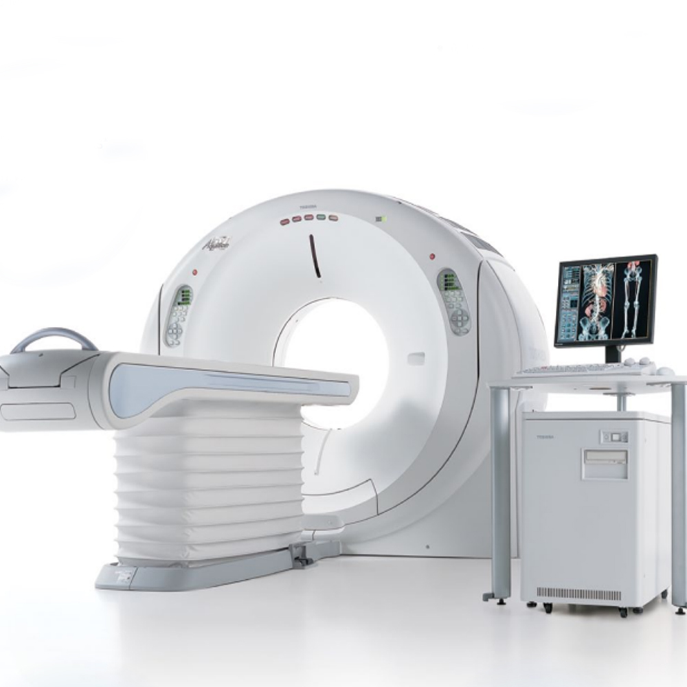
A 128-slice CT scan refers to a type of computed tomography (CT) scanner equipped with 128 detector rows, allowing for the simultaneous acquisition of multiple slices or images of the body during each rotation. This advanced technology offers several benefits over traditional CT scanners with fewer detector rows:
Faster Imaging Speed: With more detector rows, 128-slice CT scanners can acquire images of the body more rapidly compared to scanners with fewer slices. Faster imaging speed reduces examination times, which is particularly advantageous for patients who may have difficulty remaining still for long periods or for emergency cases where rapid diagnosis is critical.
Improved Spatial Resolution: The increased number of detector rows in 128-slice CT scanners allows for higher spatial resolution, meaning that smaller anatomical structures can be visualized with greater detail. Higher spatial resolution enhances the ability to detect and characterize abnormalities, improving diagnostic accuracy.
Enhanced Image Quality: The combination of faster imaging speed and improved spatial resolution results in higher-quality images with clearer delineation of anatomical structures and pathology. This enhanced image quality enables radiologists and other healthcare professionals to make more accurate diagnoses and treatment decisions.
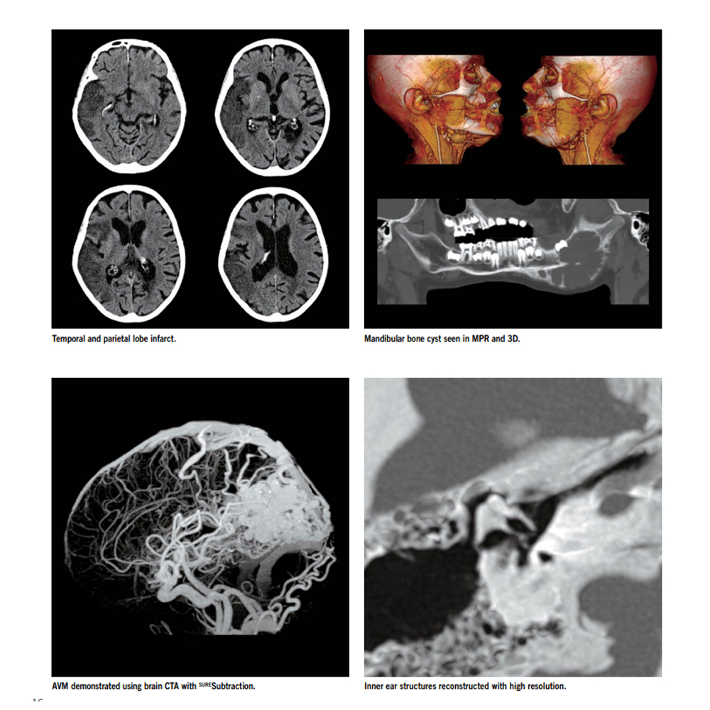
Multi-Planar Imaging and 3D Reconstruction: 128-slice CT scanners support multi-planar imaging, allowing images to be reconstructed in various planes (axial, sagittal, and coronal) without the need for additional scans.
Additionally, the acquired images can be used to generate three-dimensional (3D) reconstructions of anatomical structures, facilitating better visualization and surgical planning.
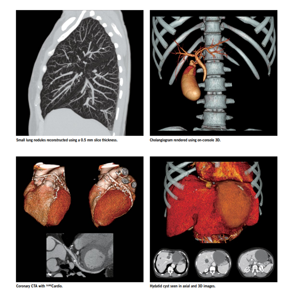
Advanced Applications: 128-slice CT scanners are capable of performing a wide range of advanced imaging techniques, including cardiac CT angiography, perfusion imaging, virtual colonoscopy, and dual-energy CT. These advanced applications provide additional diagnostic information and enable the assessment of various physiological parameters, such as blood flow and tissue perfusion.
Reduced Radiation Dose: Despite the increased number of detector rows, modern 128-slice CT scanners are designed to optimize radiation dose while maintaining image quality. Advanced dose reduction techniques, such as iterative reconstruction algorithms and dose modulation, help minimize patient exposure to ionizing radiation during imaging.
Versatility and Efficiency: 128-slice CT scanners are versatile and can accommodate a wide range of clinical applications, from routine diagnostic imaging to complex interventional procedures. Their efficiency in acquiring high-quality images in a shorter time frame contributes to improved workflow and patient throughput in healthcare settings.
In summary, 128-slice CT scanners offer several benefits, including faster imaging speed, improved spatial resolution, enhanced image quality, multi-planar imaging capabilities, advanced applications, reduced radiation dose, and versatility. These advancements contribute to more accurate diagnoses, better treatment planning, and improved patient care in various clinical scenarios.
Fast scanning times and short waiting periods are significant advantages of modern medical imaging technologies, including CT scanners with 128-slice capabilities. Here are some key points regarding these benefits:
Improved Patient Experience: Fast scanning times and short waiting periods contribute to a more positive patient experience. Patients spend less time in the scanning room, reducing discomfort and anxiety associated with prolonged procedures.
Increased Throughput: Shorter scanning times allow medical facilities to accommodate more patients within a given timeframe. This increased throughput helps reduce waiting lists and allows for more efficient use of resources.
Emergency Medicine: In emergency medicine, rapid imaging is crucial for timely diagnosis and treatment of critical conditions such as trauma, stroke, and internal bleeding. Fast scanning times enable healthcare providers to quickly assess patients and initiate appropriate interventions.
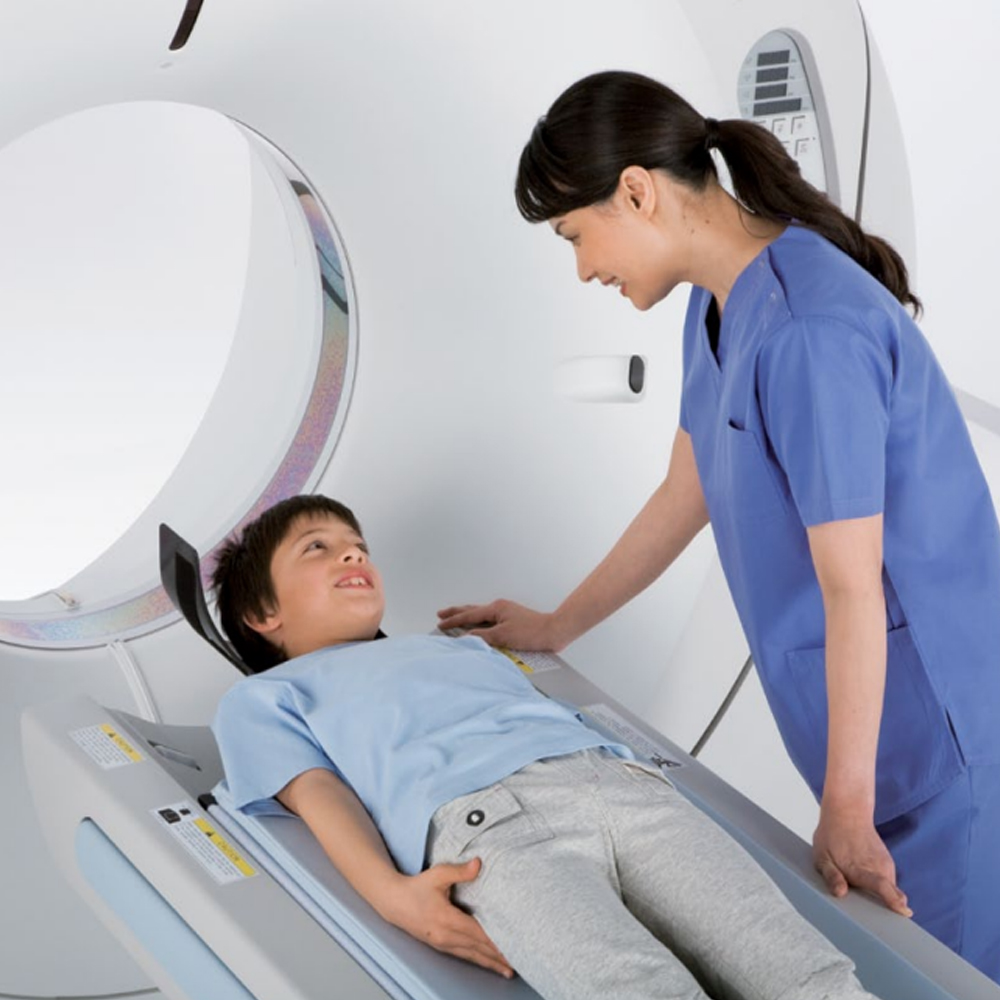
Pediatric Imaging: Short waiting periods and fast scanning times are especially beneficial for pediatric patients, who may have difficulty remaining still for extended periods. Minimizing scan time reduces the need for sedation and improves overall patient cooperation.
Enhanced Workflow: For healthcare professionals, shorter scanning times contribute to a more streamlined workflow. Radiologists can interpret images promptly, leading to faster diagnosis and treatment planning.
Outpatient Imaging Centers: In outpatient imaging centers, fast scanning times are essential for accommodating high patient volumes and maintaining efficient scheduling. Shorter waiting periods between appointments enhance patient satisfaction and improve the overall efficiency of the facility.
Reduced Radiation Exposure: Modern CT scanners are equipped with advanced technologies that optimize scanning parameters to achieve faster imaging while minimizing radiation dose. Shorter scan times contribute to reduced cumulative radiation exposure for patients undergoing multiple imaging studies over time.
Cost Efficiency: Faster scanning times translate to reduced operational costs for medical facilities. With shorter scan durations, resources such as personnel, equipment, and facility space can be utilized more efficiently, resulting in cost savings.
Overall, fast scanning times and short waiting periods offer numerous benefits for both patients and healthcare providers. These advantages improve patient comfort, enhance diagnostic capabilities, optimize resource utilization, and contribute to more efficient healthcare delivery.
Before undergoing a CT (Computed Tomography) scan, patients typically need to follow a preparation process that may include the following steps:
Medical History and Screening: Patients are usually asked to provide a detailed medical history, including any known allergies, pre-existing medical conditions, previous surgeries, and current medications. Healthcare providers may inquire about specific factors such as pregnancy, as CT scans involve exposure to ionizing radiation, which can potentially harm a developing fetus.
Instructions for Contrast Material (if applicable): If the CT scan requires the use of contrast material (contrast dye), patients may receive specific instructions regarding its administration. For oral contrast, patients may be instructed to drink a contrast agent before the scan to enhance the visibility of certain organs or structures in the abdomen or pelvis. For intravenous contrast, patients may need to fast for a few hours before the scan to ensure optimal absorption of the contrast material. Patients with allergies or a history of adverse reactions to contrast material may receive pre-medication or alternative imaging options.
Fasting (if applicable): Depending on the type of CT scan being performed, patients may be instructed to fast for a period of time before the procedure. Fasting is typically required for scans involving the abdomen or pelvis, as it helps reduce bowel gas and provides clearer images of the internal organs. Patients are advised to follow specific fasting instructions provided by their healthcare provider, including the duration of fasting and guidelines for fluid intake.
Clothing and Personal Items: Patients may be asked to change into a hospital gown before the CT scan to minimize interference from clothing containing metal components, which can affect the quality of the images. Patients should remove any metal objects, jewelry, or accessories such as belts, watches, and hairpins that may interfere with the imaging process.
Preparation for Sedation (if applicable): In some cases, patients may receive sedation before the CT scan to help them relax or remain still during the procedure. Patients receiving sedation should follow specific instructions provided by their healthcare provider regarding fasting requirements and medication administration.
Patient Education: Patients should receive detailed instructions from their healthcare provider regarding the preparation process before the CT scan. It is essential for patients to ask questions and seek clarification if they are unsure about any aspect of the preparation process or have concerns about the procedure.
Arrival Time and Check-In: Patients should arrive at the imaging facility or hospital at the designated time for their CT scan appointment. Upon arrival, patients may need to check in with the reception or registration desk and complete any necessary paperwork or consent forms before proceeding to the imaging department.
By following the preparation process outlined by their healthcare provider, patients can help ensure a smooth and successful CT scan procedure. It is essential for patients to communicate any medical conditions, allergies, or concerns they may have with their healthcare team to ensure safe and effective imaging.
Before undergoing a CT (Computed Tomography) scan, patients should take certain precautions to ensure the safety and effectiveness of the procedure. Here are some precautions to consider:
Pregnancy: Inform your healthcare provider if you are pregnant or suspect that you may be pregnant. CT scans involve exposure to ionizing radiation, which can potentially harm a developing fetus. Your healthcare provider will evaluate the necessity of the CT scan and may consider alternative imaging methods or postpone the scan until after pregnancy, if possible.
Allergies and Medical History: Inform your healthcare provider of any allergies, especially to iodine-based contrast materials commonly used in CT scans. Provide a detailed medical history, including pre-existing medical conditions, previous surgeries, and current medications.
Contrast Material Precautions: If the CT scan requires the use of contrast material (contrast dye), inform your healthcare provider if you have a history of allergic reactions to contrast agents or kidney problems. Some contrast materials contain iodine, which can cause allergic reactions in susceptible individuals or exacerbate kidney issues. Your healthcare provider may prescribe pre-medication or adjust the type and dosage of contrast material based on your medical history.
Fasting Instructions (if applicable): Follow any fasting instructions provided by your healthcare provider if the CT scan involves the abdomen or pelvis. Fasting helps reduce bowel gas and provides clearer images of the internal organs. Your healthcare provider will specify the duration of fasting and guidelines for fluid intake.
Medication Instructions: Follow your healthcare provider’s instructions regarding the use of medications before the CT scan. Depending on the type of scan and your medical history, you may need to adjust your medication schedule or temporarily stop taking certain medications.
Clothing and Personal Items: Wear comfortable clothing and remove any metal objects, jewelry, or accessories that may interfere with the imaging process. You may be asked to change into a hospital gown before the scan to minimize interference from clothing containing metal components.
Communication with Healthcare Provider: Communicate any concerns or questions you may have with your healthcare provider before the scan. Be sure to inform your healthcare provider of any changes in your health status or medication regimen since your last appointment.
By following these precautions and guidelines provided by your healthcare provider, you can help ensure a safe and successful CT scan procedure. It is essential to communicate openly with your healthcare team and address any concerns you may have before undergoing the scan.
After a CT (Computed Tomography) scan, there are several steps in the post-scan process that patients may experience:
Immediate Recovery: After the scan is complete, the patient is typically assisted off the scanning table and guided to a recovery area if needed.Patients may be monitored briefly for any immediate post-scan reactions or symptoms, especially if contrast material was used.
Resuming Normal Activities: In most cases, patients can resume their normal activities immediately after a CT scan, unless instructed otherwise by their healthcare provider. There are usually no restrictions on driving or returning to work or school following the procedure.
Contrast Material Elimination (if applicable): If contrast material was administered during the scan, it is eliminated from the body through the kidneys and excreted in the urine. Patients may be advised to drink plenty of fluids after the scan to help flush the contrast material out of their system.
Monitoring for Adverse Reactions: Patients should be aware of potential side effects or adverse reactions to contrast material, such as allergic reactions or kidney problems. Symptoms to watch for may include itching, hives, difficulty breathing, nausea, vomiting, or changes in urinary habits. Patients experiencing any concerning symptoms should contact their healthcare provider immediately.
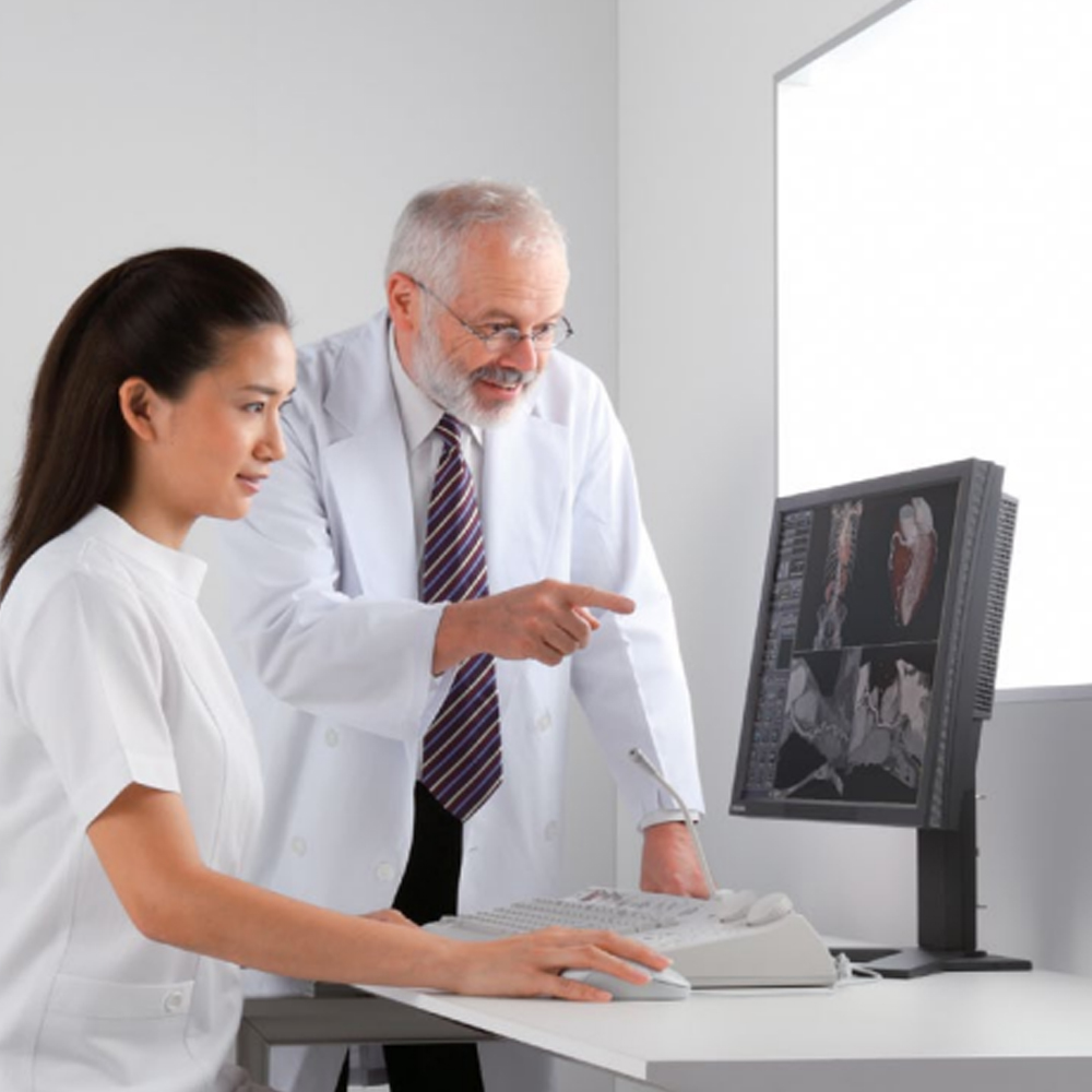
Interpreting the Results: The CT scan images are interpreted by a radiologist, who analyzes the images and prepares a report for the referring healthcare provider. Depending on the urgency of the results, patients may receive the results during a follow-up appointment with their healthcare provider, or they may be contacted by phone.
Follow-up Care: Depending on the findings of the CT scan and the reason for the imaging study, patients may require further evaluation or treatment. Healthcare providers may recommend additional tests, procedures, or consultations with specialists based on the CT scan results.
Documentation and Medical Records: The results of the CT scan, along with the radiologist’s report, are typically documented in the patient’s medical records. Patients may request a copy of the imaging report for their records or for sharing with other healthcare providers.
Patient Education: Patients may receive instructions or educational materials from their healthcare provider regarding any findings on the CT scan, follow-up care, or recommendations for managing their health condition.
Overall, the post-scan process involves monitoring for immediate reactions, resuming normal activities, monitoring for any delayed reactions, interpreting the results, and coordinating follow-up care as needed. Patients should communicate any concerns or questions they may have with their healthcare provider during this process.
Evaluation of Results and Discussion with Doctor
After a CT scan, the results are evaluated by a radiologist who specializes in interpreting diagnostic imaging studies. The radiologist carefully analyzes the images to assess anatomical structures and identify any abnormalities or findings. Based on their analysis, the radiologist prepares a detailed report summarizing the findings of the CT scan. This report is then communicated to the referring physician, who discusses the results with the patient during a follow-up appointment. During this discussion, the referring physician explains the findings of the scan, addresses any questions or concerns the patient may have, and collaborates with the patient to develop a treatment plan tailored to their individual needs. The patient is provided with education regarding the implications of the CT scan results, and a follow-up plan is established to monitor their progress and ensure continuity of care. Finally, the results of the CT scan, along with the radiologist’s report and any discussions with the doctor, are documented in the patient’s medical records for future reference.
Radiation Exposure and Safety Measures
Radiation exposure is a consideration in CT scans due to the use of ionizing radiation. However, we prioritize patient safety by implementing various measures. Our CT scanners are equipped with advanced technologies that allow for dose optimization while maintaining image quality. For instance, automatic exposure control adjusts radiation levels based on factors like patient size and tissue density. Additionally, we utilize low-dose protocols when appropriate, particularly in sensitive populations. Our staff is trained to follow strict safety protocols, including proper calibration of equipment and the use of shielding devices to minimize unnecessary exposure. Patient education is also essential; we inform patients about the benefits and risks of CT scans, including radiation exposure, and encourage open communication to address any concerns. Ultimately, our goal is to ensure that patients receive the diagnostic information they need while minimizing radiation risks as much as possible.
CT scans have a wide range of applications in various clinical conditions, offering valuable diagnostic insights and guiding treatment decisions across medical specialties. These applications include:
When undergoing a CT (Computed Tomography) scan, patients have certain rights and considerations to be aware of. These include:
Informed Consent: Patients have the right to receive comprehensive information about the purpose, risks, benefits, and alternatives of the CT scan before giving their consent for the procedure. Healthcare providers should ensure that patients understand the information provided and have the opportunity to ask questions or seek clarification.
Radiation Risks: Patients should be informed about the potential risks associated with radiation exposure from CT scans. Healthcare providers should discuss the small increased risk of cancer associated with radiation exposure, especially with repeated scans or in sensitive populations such as children and pregnant women.
Privacy and Confidentiality: Patients have the right to privacy and confidentiality regarding their medical information, including CT scan results. Healthcare providers must adhere to strict confidentiality standards and obtain patient consent before sharing medical information with third parties.
Patient Comfort and Safety: Patients should receive appropriate care to ensure their comfort and safety during the CT scan procedure. Healthcare providers should address patient concerns, provide support, and ensure that the scan is conducted in a safe and professional manner.
Access to Information: Patients have the right to access their medical records, including CT scan results, and to receive explanations and interpretations of the findings from their healthcare providers. Open communication and transparency regarding the CT scan results are essential to empower patients in their healthcare decisions.
Trauma and Emergency Medicine: CT scans play a crucial role in evaluating traumatic injuries such as head trauma, spinal injuries, and fractures. They provide detailed imaging of the affected areas, aiding in the rapid diagnosis and treatment of life-threatening conditions.
Oncology (Cancer Imaging): CT scans are essential in cancer diagnosis, staging, and treatment planning. They help identify tumors, determine their size, location, and extent, and assess their response to treatment. CT scans are particularly useful in detecting solid tumors in organs such as the lungs, liver, and abdomen.
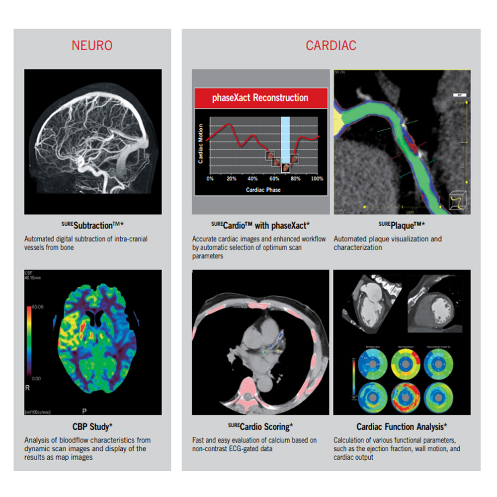
Neurological Disorders: In neurology, CT scans are utilized to evaluate various conditions affecting the brain and spine, including strokes, brain tumors, and degenerative diseases. They provide detailed imaging of brain structures, aiding in the diagnosis and management of neurological disorders.
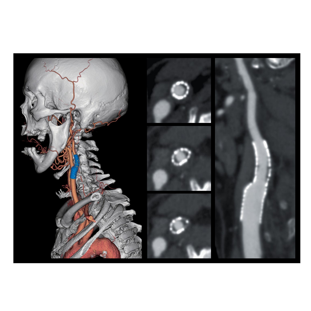
Cardiovascular Medicine: CT angiography is used in cardiovascular medicine to assess heart and vascular diseases. It provides detailed images of the heart and blood vessels, helping diagnose conditions such as coronary artery disease, aortic aneurysms, and pulmonary embolism.
Orthopedics and Musculoskeletal Disorders: CT scans are valuable in evaluating musculoskeletal injuries and disorders, including fractures, joint abnormalities, and bone tumors. They provide detailed images of bones and soft tissues, aiding orthopedic surgeons in treatment planning and surgical interventions.
Abdominal and Pelvic Conditions: CT scans are commonly used to assess abdominal and pelvic conditions such as gastrointestinal disorders, kidney stones, and reproductive organ abnormalities. They provide detailed imaging of internal organs, facilitating the diagnosis and management of various abdominal and pelvic disorders.
Pulmonary Medicine: CT scans are instrumental in evaluating pulmonary conditions such as lung nodules, pneumonia, and chronic obstructive pulmonary disease (COPD). They provide detailed imaging of the lungs and airways, aiding pulmonologists in diagnosis and treatment planning.
Overall, CT scans are versatile imaging modalities with widespread applications across medical specialties, providing valuable diagnostic information and guiding treatment decisions in a variety of clinical conditions.
Privacy and data protection are essential considerations in the context of medical imaging, including CT (Computed Tomography) scans. Patients’ medical information, including imaging results, must be handled with strict confidentiality and protected from unauthorized access or disclosure. Healthcare providers and facilities must adhere to legal and ethical standards to ensure patient privacy and data security. Measures to safeguard privacy and data protection in CT scans include:
Confidentiality: Patient medical information, including CT scan results, should be treated with the utmost confidentiality. Healthcare providers must ensure that only authorized personnel have access to patient records, and patient information should not be shared with third parties without the patient’s consent.
Secure Storage: CT scan images and related medical records should be stored securely in electronic health record systems or physical files. Access to patient data should be restricted to authorized personnel through password-protected systems or other secure means.
Data Encryption: Electronic transmission of patient data, including CT scan images, should be encrypted to protect against unauthorized interception or access. Encryption helps safeguard patient privacy and prevents data breaches during transmission over networks.
Access Controls: Healthcare facilities should implement access controls to limit access to patient data based on the principle of least privilege. Only authorized personnel with a legitimate need to access patient information should be granted permission to do so.
Training and Awareness: Healthcare staff should receive training on privacy and data protection practices, including handling patient information securely and adhering to confidentiality protocols. Regular awareness programs can help reinforce the importance of privacy and data security among healthcare professionals.
Compliance with Regulations: Healthcare providers and facilities must comply with relevant regulations and standards related to patient privacy and data protection, such as the Health Insurance Portability and Accountability Act (HIPAA) in the United States or the General Data Protection Regulation (GDPR) in the European Union. Compliance with these regulations ensures that patient data is handled ethically and legally.
Data Retention Policies: Healthcare facilities should establish data retention policies that specify the length of time patient data, including CT scan images, will be retained. Retention periods should be based on legal requirements and clinical needs, and data should be securely disposed of once it is no longer needed.
By implementing these measures, healthcare providers and facilities can ensure that patient privacy and data protection are upheld in the context of CT scans and other medical imaging procedures. This helps build trust with patients and ensures that their sensitive medical information is handled securely and confidentially.
During the scanning process, patients have specific rights and preferences that should be respected to ensure a positive and comfortable experience. These include:
Informed Consent: Patients have the right to receive clear and understandable information about the CT scanning procedure, including its purpose, benefits, potential risks, and alternatives. They should be given the opportunity to ask questions and express any concerns before providing their consent for the scan.
Privacy and Dignity: Patients should be treated with respect for their privacy and dignity during the scanning process. Healthcare providers should ensure that patients are provided with appropriate clothing or coverings to maintain their privacy while undergoing the scan.
Comfort and Safety: Patients have the right to feel comfortable and safe during the scanning process. Healthcare providers should take measures to address any discomfort or anxiety experienced by patients, such as providing support, explaining the procedure in detail, and ensuring that the scan is conducted in a safe and professional manner.

 Multi-Slice Computed Tomography (CT), also known as Multi-Slice CT, is a medical imaging technique that utilizes X-rays to generate detailed cross-sectional images of the body. The term “128 slices” refers to the number of detectors or slices present in the CT scanner. Here’s a description of CT scanning and its components: