Burtom ‣ Technologies ‣ Computed Tomography 384 Slices (Multi-Slice CT)
Language: 🇬🇧 English | 🇹🇷 Türkçe
Computed Tomography 384 Slices (Multi-Slice CT) Overview

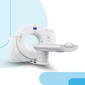 Multi-Slice CT technology has revolutionized diagnostic imaging, and the latest iteration with 384 slices offers unparalleled capabilities. By capturing multiple slices of the body simultaneously, this advanced CT scanner provides detailed cross-sectional images with exceptional clarity and precision. The high slice count enables rapid scanning of large anatomical areas, resulting in faster imaging times and improved patient throughput.
With its increased spatial resolution and enhanced image quality, Multi-Slice CT 384 Slices delivers superior diagnostic accuracy across a wide range of medical applications. From detecting subtle abnormalities in the brain and vascular structures to evaluating complex fractures and assessing tumor response to treatment, this advanced imaging modality empowers healthcare providers with invaluable clinical information.
Furthermore, Multi-Slice CT with 384 slices enables advanced imaging techniques such as perfusion imaging, dual-energy CT, and cardiac imaging with reduced radiation exposure and contrast dose. Its versatility and advanced capabilities make it an indispensable tool in modern medical practice, facilitating precise diagnosis, treatment planning, and post-treatment monitoring.
In summary, Computed Tomography 384 Slices (Multi-Slice CT) represents the pinnacle of diagnostic imaging technology, offering unparalleled image quality, speed, and clinical utility.
Multi-Slice CT technology has revolutionized diagnostic imaging, and the latest iteration with 384 slices offers unparalleled capabilities. By capturing multiple slices of the body simultaneously, this advanced CT scanner provides detailed cross-sectional images with exceptional clarity and precision. The high slice count enables rapid scanning of large anatomical areas, resulting in faster imaging times and improved patient throughput.
With its increased spatial resolution and enhanced image quality, Multi-Slice CT 384 Slices delivers superior diagnostic accuracy across a wide range of medical applications. From detecting subtle abnormalities in the brain and vascular structures to evaluating complex fractures and assessing tumor response to treatment, this advanced imaging modality empowers healthcare providers with invaluable clinical information.
Furthermore, Multi-Slice CT with 384 slices enables advanced imaging techniques such as perfusion imaging, dual-energy CT, and cardiac imaging with reduced radiation exposure and contrast dose. Its versatility and advanced capabilities make it an indispensable tool in modern medical practice, facilitating precise diagnosis, treatment planning, and post-treatment monitoring.
In summary, Computed Tomography 384 Slices (Multi-Slice CT) represents the pinnacle of diagnostic imaging technology, offering unparalleled image quality, speed, and clinical utility. 


What is a CT Scan and How is it Performed?
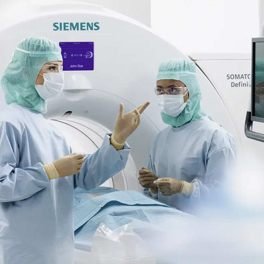
A CT scan, also known as computed tomography scan, is a medical imaging technique that uses X-rays and computer processing to create detailed cross-sectional images of the body. During a CT scan, the patient lies on a motorized table that moves through a doughnut-shaped machine called a CT scanner. The scanner emits X-rays from multiple angles around the body, and detectors measure the amount of radiation that passes through the body. The data collected is processed by a computer to create detailed cross-sectional images, which can be viewed on a monitor and further analyzed by healthcare professionals. CT scans are commonly used to diagnose and monitor various medical conditions, including injuries, tumors, infections, and internal bleeding.
Effects of CT Scan on the Patient
CT scans use X-rays, which involve ionizing radiation, to create detailed images of the body’s internal structures. While CT scans are generally safe and widely used in medical practice, they do expose patients to a small amount of radiation. The radiation exposure from a single CT scan is typically low and usually does not cause any immediate harmful effects. However, repeated exposure to radiation from multiple CT scans over time may increase the risk of developing certain radiation-related health issues, such as cancer.
It’s essential for healthcare providers to weigh the benefits of obtaining diagnostic information from a CT scan against the potential risks associated with radiation exposure, especially for children, pregnant women, and individuals with underlying health conditions. In some cases, alternative imaging modalities that do not involve ionizing radiation, such as MRI or ultrasound, may be considered to minimize radiation exposure.
To mitigate the risks associated with radiation exposure during CT scans, healthcare providers adhere to established guidelines and protocols to ensure that scans are performed only when necessary and that radiation doses are kept as low as reasonably achievable (ALARA). Additionally, patients undergoing CT scans are typically provided with protective shielding, and healthcare professionals carefully monitor and optimize scanning parameters to minimize radiation exposure while obtaining diagnostic images of sufficient quality.
Patient Experience During CT Scan
The patient experience during a CT scan can vary depending on several factors, including the reason for the scan, the individual’s medical condition, and their personal preferences. Here are some common aspects of the patient experience during a CT scan:
Preparation: Before the CT scan, patients may be asked to change into a hospital gown and remove any metal objects or jewelry that could interfere with the scan. They may also need to follow specific instructions, such as fasting for a certain period or abstaining from certain medications.
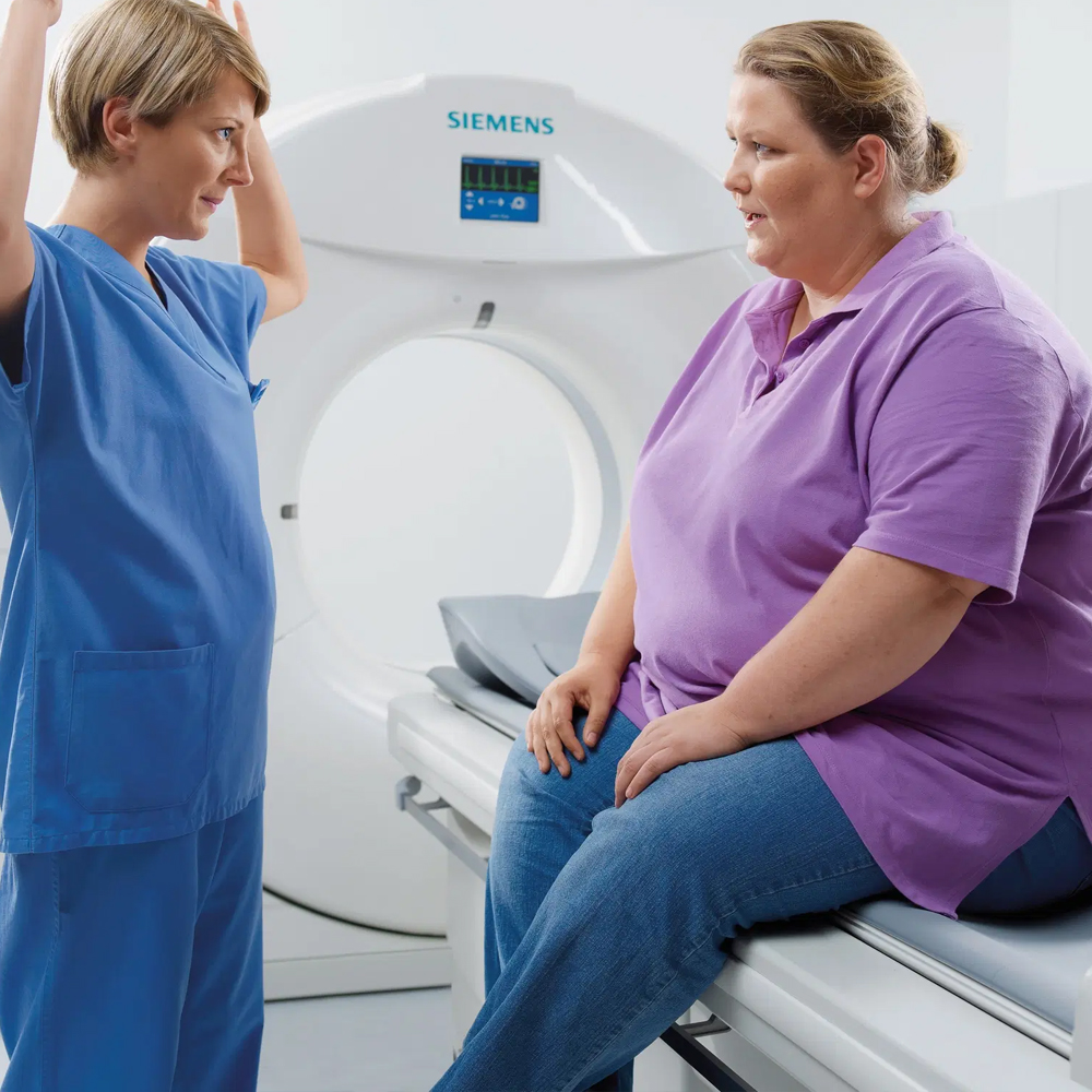
Positioning: During the scan, the patient lies on a motorized table that moves through the CT scanner. The technologist helps position the patient comfortably on the table and may use pillows or cushions for support.
Immobility: It’s essential for patients to remain still during the CT scan to ensure clear and accurate images. Depending on the type of scan and the area being examined, patients may need to hold their breath briefly or remain in a specific position for a short period.
Contrast dye: In some cases, a contrast dye may be administered intravenously to enhance the visibility of certain tissues or organs during the scan. Patients may experience a warm sensation or a metallic taste in their mouth when the contrast dye is injected.
Noise and motion: CT scanners emit buzzing or whirring noises during the scan, which can be loud and may cause some discomfort for some patients. The table may also move slowly through the scanner during the imaging process.
Duration: The duration of a CT scan can vary depending on the type of scan and the area being examined. Some scans may be completed within a few minutes, while others may take longer.
Communication: Patients are typically in constant communication with the technologist operating the CT scanner through an intercom system. They are informed about the steps of the procedure and may be asked to provide feedback or follow specific instructions during the scan.
Comfort measures: Healthcare providers strive to ensure the patient’s comfort and well-being throughout the procedure. Patients are encouraged to communicate any discomfort or concerns they may have, and adjustments can be made as needed to enhance their comfort during the scan.
Overall, while undergoing a CT scan may be an unfamiliar experience for some patients, healthcare providers strive to ensure that patients are informed, comfortable, and well-supported throughout the procedure.
384-Slice CT Scan and Its Benefits
A 384-slice CT scan, also known as a multi-slice CT scan, is an advanced imaging technique that provides high-resolution images of the body using multiple thin slices. Here are some of the benefits of a 384-slice CT scan:
Improved Image Quality: With more slices, a 384-slice CT scanner can produce higher resolution images with greater detail compared to lower slice scanners. This enhanced image quality allows for better visualization of structures and abnormalities within the body.
Faster Scanning Speed: The increased number of slices allows for faster scanning times, reducing the time required for the entire imaging process. This is particularly beneficial for patients who may have difficulty remaining still during the scan or for those who are unable to tolerate longer scan times.
Enhanced Diagnostic Accuracy: The high-resolution images produced by a 384-slice CT scan enable healthcare providers to make more accurate diagnoses and treatment plans. The detailed images allow for better visualization of small lesions, tumors, or other abnormalities that may not be easily detected with lower slice scanners.
Reduced Radiation Dose: Despite the increased number of slices, advancements in CT technology have allowed for the development of dose-reduction techniques. This means that patients undergoing a 384-slice CT scan may be exposed to lower levels of radiation compared to older CT scanners, while still achieving high-quality images.
Comprehensive Imaging Capabilities: A 384-slice CT scanner can perform a wide range of imaging studies, including angiography, cardiac imaging, and whole-body scans. This versatility allows healthcare providers to assess various anatomical structures and conditions in detail, contributing to more comprehensive patient care.
Improved Patient Experience: The faster scanning times and higher image quality of a 384-slice CT scan contribute to a more comfortable and efficient patient experience. Shorter scan times reduce the need for patients to hold their breath or remain still for extended periods, leading to less discomfort and anxiety during the procedure.
Overall, the 384-slice CT scan offers numerous benefits in terms of image quality, speed, accuracy, radiation dose reduction, and patient experience, making it a valuable tool for diagnostic imaging in healthcare settings.
High Resolution and Detailed Imaging
High-resolution and detailed imaging refer to the quality and clarity of the images produced by a medical imaging technique, such as a CT scan or MRI. These images provide healthcare professionals with a clear view of the structures and tissues within the body, allowing for more accurate diagnoses and treatment planning.
The term “high resolution” indicates that the images have a high level of detail, meaning that even small structures or abnormalities can be visualized with clarity. This is important for detecting subtle changes or abnormalities that may be indicative of disease or injury.
Detailed imaging refers to the ability of the imaging technique to capture fine anatomical features and differentiate between different types of tissues. For example, in CT scans, detailed imaging can distinguish between organs, blood vessels, bones, and soft tissues, providing a comprehensive view of the internal anatomy.
High-resolution and detailed imaging are essential in various medical applications, including diagnosing conditions such as cancer, cardiovascular disease, neurological disorders, and musculoskeletal injuries. By providing clear and detailed images, these imaging techniques help healthcare professionals make more informed decisions about patient care and treatment.
Fast Scanning Times and Short Waiting Periods
Fast scanning times and short waiting periods refer to the efficiency of the imaging process in medical procedures such as CT scans or MRI. These terms highlight the speed at which images are acquired and the minimal time patients need to wait during the scanning process.
Fast scanning times indicate the duration it takes for the imaging equipment to capture the necessary images of the patient’s body. This efficiency is crucial in medical settings where timely diagnosis and treatment are critical. Short waiting periods refer to the minimal time patients spend waiting before and during the imaging procedure, including scheduling, preparation, and actual scanning time.
Reducing scanning times and waiting periods improves patient satisfaction, increases throughput in medical facilities, and enhances overall workflow efficiency. It also minimizes patient discomfort and anxiety associated with prolonged waiting times, contributing to a more positive healthcare experience. Additionally, faster scanning times and shorter waiting periods allow medical professionals to accommodate more patients, leading to improved access to diagnostic services and timely medical interventions.
Preparation Process Before CT Scan
The preparation process before a CT scan involves several steps to ensure accurate imaging and patient safety. Here are some common aspects of the preparation process:
Medical History and Screening: Patients may be asked to provide information about their medical history, allergies, medications, and any previous imaging studies. It’s essential to inform the healthcare team about any existing medical conditions or concerns.
Contrast Agent Administration: In some cases, a contrast agent may be administered to enhance the visibility of certain tissues or organs during the CT scan. Patients may need to fast for a few hours before receiving the contrast agent, depending on the type of scan and medical history.
Clothing and Jewelry Removal: Patients may be asked to change into a hospital gown and remove any clothing or jewelry that could interfere with the imaging process. Metal objects such as jewelry, eyeglasses, or hearing aids can disrupt the CT scan and should be left outside the scanning room.
Patient Positioning: Depending on the area of the body being scanned, patients may need to lie down on a scanning table in a specific position. The healthcare team will provide instructions to ensure proper positioning for accurate imaging.
Breathing and Motion Instructions: Patients may be instructed to hold their breath or remain still during certain parts of the scan to minimize motion artifacts and ensure clear images. It’s essential to follow these instructions carefully to obtain high-quality scans.
Communication with the Healthcare Team: Patients should communicate any concerns or questions they have about the procedure with the healthcare team. It’s essential to address any anxieties or discomfort beforehand to ensure a smooth and successful imaging experience.
Overall, the preparation process before a CT scan aims to optimize imaging quality, ensure patient safety, and provide a comfortable experience for the individual undergoing the procedure. Following pre-scan instructions and communicating openly with the healthcare team can help facilitate a successful CT scan.
Precautions Before the Scan
Before undergoing a CT scan, patients may need to take certain precautions to ensure the procedure is safe and effective. Here are some common precautions:
Informing the Healthcare Team: Patients should inform their healthcare provider about any allergies, medical conditions, or medications they are taking, especially if they have a history of allergic reactions to contrast agents or iodine.
Fasting: Depending on the type of CT scan being performed, patients may need to fast for a period before the procedure. This is typically required for scans that involve the use of contrast agents to enhance imaging results.
Contrast Agent Precautions: If contrast dye is going to be used during the CT scan, patients should inform their healthcare provider if they have any kidney problems, as contrast agents can affect kidney function. Additionally, patients with a history of contrast dye reactions should inform their healthcare provider.
Pregnancy and Radiation Exposure: Pregnant women should inform their healthcare provider before undergoing a CT scan, as the procedure involves exposure to ionizing radiation which may pose risks to the developing fetus. In some cases, alternative imaging methods that do not use radiation may be considered for pregnant women.
Clothing and Jewelry: Patients may be asked to remove clothing and jewelry that could interfere with the imaging process, such as metal objects that may create artifacts on the images. Hospital gowns are usually provided for the scan.
Patient Positioning: Patients should follow instructions from the healthcare team regarding positioning on the CT scanner table to ensure optimal imaging results.
Communication: Patients should communicate any concerns or questions they have about the procedure with the healthcare team. It’s essential to address any anxieties or discomfort beforehand to ensure a smooth and successful imaging experience.
Following these precautions can help ensure the safety and effectiveness of the CT scan procedure. It’s important for patients to discuss any specific concerns or considerations with their healthcare provider before the scan.
Tips for Enhancing Patient Satisfaction
Enhancing patient satisfaction during a CT scan involves providing a supportive and comfortable environment while addressing patients’ needs and concerns. Here are some tips for improving patient satisfaction:
Clear Communication: Ensure clear and effective communication with patients throughout the entire process. Explain the procedure in detail, including what to expect during the scan, how long it will take, and any potential sensations they may experience.
Empathy and Compassion: Show empathy and compassion towards patients’ concerns and fears. Acknowledge their feelings and provide reassurance throughout the procedure.
Comfortable Environment: Create a comfortable and calming environment in the CT scanning room. Consider factors such as lighting, temperature, and noise level to help patients feel more relaxed during the procedure.
Privacy and Dignity: Respect patients’ privacy and dignity by providing appropriate gowning and ensuring confidentiality during discussions.
Patient Education: Educate patients about the importance of the CT scan and how it will benefit their healthcare. Answer any questions they may have and address any misconceptions about the procedure.
Minimize Wait Times: Minimize wait times for appointments and during the scanning process to reduce patient anxiety and frustration.
Pain Management: Offer pain management techniques or medications if needed to help alleviate any discomfort or anxiety associated with the procedure.
Follow-up and Support: Provide follow-up support after the CT scan, including discussing the results with the patient and addressing any additional questions or concerns they may have.
Feedback Mechanism: Implement a feedback mechanism to gather patients’ opinions and suggestions for improving the CT scanning experience. Use this feedback to make continuous improvements in patient care.
Empowerment: Empower patients by involving them in decision-making processes related to their care and providing them with information and resources to make informed choices.
By incorporating these tips into the CT scanning process, healthcare providers can help enhance patient satisfaction and improve overall patient experience.
Patient Comfort During CT Scan
Ensuring patient comfort during a CT scan is crucial for a positive experience. Here are some strategies to enhance patient comfort during the procedure:
Clear Communication: Explain the CT scan procedure to the patient in detail beforehand, including what to expect during the scan, how long it will take, and any sensations they may experience.
Comfortable Positioning: Position the patient comfortably on the CT scanning table, ensuring they are relaxed and supported throughout the procedure.
Minimal Discomfort: Minimize discomfort during positioning by using supportive cushions or pads, especially for patients with mobility issues or discomfort in specific body areas.
Warmth and Privacy: Maintain a warm and private environment in the CT scanning room to help patients feel more comfortable and at ease.
Anxiety Reduction: Address patient anxiety by providing reassurance, answering questions, and offering relaxation techniques such as deep breathing exercises or calming music.
Claustrophobia Management: For patients with claustrophobia, consider using open CT scanners or providing relaxation techniques to help them feel more comfortable during the procedure.
Pain Management: Manage any pain or discomfort experienced by the patient during the scan by offering pain-relief options or adjusting positioning as needed.
Continuous Monitoring: Continuously monitor the patient during the scan to ensure their comfort and well-being, and be responsive to any discomfort or concerns they may express.
Empowerment: Empower the patient by involving them in the process, allowing them to ask questions, express concerns, and participate in decisions related to their care.
Post-Scan Support: Offer post-scan support and assistance, including discussing the results with the patient and addressing any further questions or concerns they may have.
By prioritizing patient comfort and employing these strategies, healthcare providers can help create a more positive and comfortable CT scanning experience for patients.
Comfortable Positioning and Remaining Still
Ensuring comfortable positioning and maintaining stillness during a CT scan are essential for obtaining clear and accurate images while minimizing patient discomfort. Here are some strategies to achieve this:
Proper Positioning: Position the patient comfortably on the CT scanning table, ensuring adequate support for the head, neck, and body. Use positioning aids such as cushions or foam pads to maintain a comfortable posture.
Clear Instructions: Provide clear instructions to the patient regarding the desired position and the importance of remaining still during the scan. Explain the procedure and reassure the patient about the duration of the scan.
Supportive Equipment: Utilize positioning aids and immobilization devices, such as straps or foam pads, to help the patient maintain the required position and reduce movement during the scan.
Communication: Maintain open communication with the patient throughout the procedure. Encourage them to communicate any discomfort or need for adjustment during the scan.
Patient Education: Educate the patient about the importance of remaining still during the scan to obtain clear images. Emphasize the role of their cooperation in ensuring an accurate diagnosis.
Preparation: Prepare the patient physically and mentally for the scan by providing relaxation techniques or breathing exercises to help them remain calm and still during the procedure.
Minimize Discomfort: Address any potential sources of discomfort before the scan, such as positioning-related pressure points or claustrophobia. Adjust the positioning or offer additional support as needed to enhance comfort.
Monitoring: Continuously monitor the patient’s position and movement during the scan. Use real-time imaging or feedback systems to detect and correct any deviations from the desired position.
Patient Engagement: Engage the patient in the process by encouraging them to actively participate in maintaining stillness. Offer positive reinforcement and encouragement throughout the scan.
Posture Coaching: Provide coaching on proper posture and relaxation techniques before the scan to help the patient maintain a comfortable and still position during the procedure.
By implementing these strategies, healthcare providers can help ensure comfortable positioning and encourage patients to remain still during CT scans, leading to improved image quality and a more positive patient experience.
Post-Scan Process
After the CT scan is completed, there are several steps involved in the post-scan process to ensure proper follow-up and patient care:
Image Review: The images obtained from the CT scan are reviewed by a radiologist or a qualified medical professional to evaluate the structures and areas of interest. The radiologist interprets the images to make a diagnosis or assess the condition being evaluated.
Results Communication: The results of the CT scan are communicated to the referring physician or healthcare provider who ordered the scan. The physician discusses the findings with the patient and explains the implications of the results for their diagnosis and treatment plan.
Treatment Planning: Based on the findings of the CT scan, the healthcare provider develops a treatment plan tailored to the patient’s condition. This may involve medication, further diagnostic tests, surgical intervention, or other forms of treatment as deemed necessary.
Patient Education: The patient is educated about the results of the CT scan, including any abnormalities detected and the recommended course of action. They are provided with information about their condition, treatment options, potential risks, and expected outcomes.
Follow-up Care: Patients may require follow-up appointments with their healthcare provider to monitor their condition, assess treatment effectiveness, and adjust the treatment plan if needed. Follow-up imaging studies may also be scheduled to track changes over time.
Documentation: The results of the CT scan and related clinical information are documented in the patient’s medical record for future reference. This ensures that all relevant information is readily available to healthcare providers involved in the patient’s care.
Patient Support: Patients may have questions or concerns about the results of their CT scan and the recommended treatment plan. Healthcare providers offer support, address their concerns, and provide guidance to help them navigate their healthcare journey.
Billing and Administrative Tasks: Administrative tasks, such as billing for the CT scan and processing insurance claims, are completed according to established protocols. Patients may receive information about billing and payment options as needed.
Continued Monitoring: For certain conditions or treatments, ongoing monitoring and follow-up may be necessary to track progress and ensure optimal outcomes. Patients are advised on the importance of adhering to follow-up appointments and recommended care guidelines.
By effectively managing the post-scan process, healthcare providers ensure seamless continuity of care for patients undergoing CT scans, leading to timely diagnosis, appropriate treatment, and improved patient outcomes.
Evaluation of Results and Discussion with Doctor
After the completion of a CT scan, the results are evaluated by a radiologist or a qualified medical professional. The evaluation process involves carefully reviewing the images obtained during the scan to assess the structures and areas of interest in the body. The radiologist interprets the images to identify any abnormalities, lesions, or other findings that may be relevant to the patient’s condition.
Once the results have been evaluated, the radiologist communicates their findings to the referring physician or healthcare provider who ordered the CT scan. This communication typically involves providing a detailed report summarizing the findings and their clinical significance.
Following the evaluation of results, a discussion takes place between the patient and their doctor. During this discussion, the doctor explains the findings of the CT scan to the patient in a clear and understandable manner. They discuss any abnormalities detected, their implications for the patient’s health, and the recommended course of action.
The discussion with the doctor may cover various aspects, including:
Diagnosis: The doctor explains the diagnosis based on the CT scan findings, if applicable. They provide information about the condition being evaluated and its potential implications for the patient’s health.
Treatment Options: Based on the CT scan results, the doctor discusses the available treatment options with the patient. This may include medications, lifestyle modifications, further diagnostic tests, or other interventions.
Prognosis: The doctor discusses the prognosis or outlook for the patient’s condition based on the CT scan findings. They explain the expected course of the condition and the potential outcomes of treatment.
Follow-up Care: The doctor outlines the recommended follow-up care plan, which may include additional appointments, monitoring, or further testing. They explain the importance of follow-up care in managing the patient’s condition effectively.
Patient Questions and Concerns: During the discussion, the patient is encouraged to ask questions and express any concerns they may have about the CT scan results or their condition. The doctor provides clarification and addresses any issues raised by the patient.
Patient Education: The doctor educates the patient about their condition, treatment options, and steps they can take to manage their health effectively. They provide information to empower the patient to make informed decisions about their care.
Overall, the evaluation of CT scan results and discussion with the doctor play a crucial role in ensuring that patients understand their health status, are actively involved in decision-making regarding their care, and receive appropriate guidance and support from their healthcare provider.
Risks and Safety of 384-Slice CT Scan
The safety of a 384-slice CT scan, like any medical procedure involving radiation, is a topic of concern for patients and healthcare providers. While CT scans are generally considered safe and beneficial for diagnosing various medical conditions, they do involve exposure to ionizing radiation, which carries certain risks. Here are some considerations regarding the risks and safety of 384-slice CT scans:
Radiation Exposure: CT scans use X-rays to create detailed cross-sectional images of the body. The amount of radiation exposure during a CT scan is higher compared to other imaging modalities, such as X-rays or MRI. With a 384-slice CT scan, the radiation dose may be higher due to the increased number of detector rows and the need for more image acquisitions.
Potential Risks: The main risk associated with radiation exposure from CT scans is the theoretical increased risk of developing cancer later in life. This risk is generally considered low, especially for single CT scans. However, repeated or unnecessary CT scans over time may increase the cumulative radiation dose and consequently the potential risk of cancer.
Risk-Benefit Analysis: The decision to perform a CT scan, including a 384-slice CT scan, involves a risk-benefit analysis. Healthcare providers weigh the potential benefits of obtaining detailed diagnostic information against the potential risks of radiation exposure. In many cases, the benefits of accurate diagnosis and appropriate medical management outweigh the small potential risks associated with radiation exposure.
Radiation Dose Reduction Techniques: Radiology departments employ various techniques to minimize radiation dose during CT scans. These include using lower radiation settings, optimizing scan protocols, and employing dose modulation techniques based on patient size and anatomy. Additionally, newer CT technologies, such as iterative reconstruction algorithms, help reduce radiation dose while maintaining image quality.
Patient Safety Measures: Healthcare providers take several steps to ensure patient safety during CT scans. This includes screening patients for contraindications, such as pregnancy, and obtaining informed consent. Radiology technologists also ensure that the CT scanner is properly calibrated and that patients are positioned correctly to minimize radiation exposure.
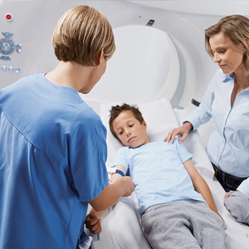
Pediatric Considerations: Special considerations apply to pediatric patients undergoing CT scans, as they are more sensitive to radiation. Radiology departments follow pediatric imaging guidelines and use dose reduction techniques to minimize radiation exposure while maintaining diagnostic image quality.
In summary, while 384-slice CT scans offer advanced imaging capabilities, it’s essential to consider the potential risks of radiation exposure and take appropriate measures to ensure patient safety. Healthcare providers strive to balance the benefits of diagnostic imaging with the risks, employing dose reduction techniques and adhering to best practices to minimize radiation exposure during CT scans.
Patient Safety During the Scan
Ensuring patient safety during a CT scan, including a 384-slice CT scan, is a critical aspect of the imaging process. Here are some key considerations for maintaining patient safety during the scan:
Patient Screening: Before the CT scan, healthcare providers screen patients for any contraindications or factors that may affect the safety of the procedure. This includes assessing for allergies, medical conditions, and pregnancy, as well as obtaining a complete medical history.
Informed Consent: Patients should receive clear information about the CT scan procedure, including its purpose, benefits, risks, and alternatives. Informed consent is obtained from the patient or their legal guardian before the scan to ensure they understand and agree to the procedure.
Radiation Safety: Radiology technologists ensure that the CT scanner is properly calibrated and that radiation dose levels are optimized for each patient. Techniques such as automatic exposure control and dose modulation are used to adjust radiation dose based on patient size and anatomy while maintaining image quality.
Patient Positioning: Proper patient positioning is essential for obtaining accurate CT images and minimizing radiation exposure. Patients are positioned carefully on the CT table, and immobilization devices may be used to ensure they remain still during the scan.
Monitoring and Communication: Radiology technologists monitor patients throughout the scan and maintain clear communication with them via intercom or visual contact. Patients are informed about the duration of the scan and any instructions they need to follow, such as holding their breath during specific image acquisitions.
Emergency Preparedness: Radiology departments have protocols in place for handling emergency situations during CT scans. This includes immediate access to emergency equipment, such as crash carts and defibrillators, as well as trained staff who can respond quickly to any medical emergencies.
Pediatric Considerations: Special attention is given to pediatric patients undergoing CT scans to ensure their safety. Pediatric imaging protocols are used to minimize radiation dose while obtaining diagnostic images. Child-friendly approaches, such as distraction techniques or sedation when necessary, may also be employed to help young patients feel more comfortable during the scan.
Overall, patient safety is paramount during CT scans, and healthcare providers follow stringent protocols to minimize risks and ensure a safe and effective imaging experience for every patient.
Applications of CT Scan in Various Clinical Conditions
CT scans, including 384-slice CT scans, are versatile imaging tools used in various clinical conditions across different medical specialties. Here are some common applications of CT scans in clinical practice:
Trauma and Emergency Medicine: CT scans are valuable in assessing traumatic injuries, such as fractures, internal bleeding, and organ damage, providing rapid and detailed imaging to guide emergency management.
Neurology: CT scans of the brain are used to evaluate stroke, hemorrhage, brain tumors, and other neurological conditions. CT angiography (CTA) can assess blood vessels in the brain, aiding in the diagnosis of conditions like aneurysms and arteriovenous malformations.
Oncology: CT scans are essential for diagnosing and staging various cancers, including lung cancer, liver cancer, pancreatic cancer, and lymphomas. They help detect tumors, assess tumor size and location, monitor treatment response, and detect metastases.
Cardiology: CT angiography (CTA) is used to evaluate the heart and blood vessels, detecting coronary artery disease, assessing cardiac function, and evaluating congenital heart defects. Cardiac CT is also used in planning for interventions like coronary artery bypass grafting (CABG) and transcatheter aortic valve replacement (TAVR).
Pulmonology: CT scans of the chest are crucial for diagnosing and monitoring respiratory conditions such as pneumonia, pulmonary embolism, chronic obstructive pulmonary disease (COPD), and lung nodules. They are also used for lung cancer screening in high-risk individuals.
Gastroenterology: CT scans are used to evaluate the gastrointestinal tract, including the liver, pancreas, gallbladder, and intestines. They help diagnose conditions like liver cirrhosis, pancreatic cancer, gallstones, and inflammatory bowel disease (IBD).
Orthopedics: CT scans provide detailed imaging of bones and joints, aiding in the diagnosis and management of orthopedic conditions such as fractures, arthritis, spinal disorders, and joint abnormalities.
Urology: CT scans of the abdomen and pelvis are used to evaluate the urinary tract, kidneys, bladder, and reproductive organs. They assist in diagnosing conditions like kidney stones, urinary tract infections, renal masses, and prostate cancer.
Vascular Imaging: CT angiography (CTA) is widely used to visualize blood vessels throughout the body, helping diagnose vascular diseases such as aortic aneurysms, peripheral artery disease (PAD), and carotid artery stenosis.
Pediatrics: CT scans are used in pediatric patients to evaluate congenital anomalies, assess traumatic injuries, diagnose infections, and monitor the progression of diseases.
These are just a few examples of the many clinical applications of CT scans, highlighting their versatility and importance in modern medical practice across various specialties.
Patient Rights and Important Considerations Regarding CT Scan
Patients undergoing CT scans have certain rights and considerations that should be respected to ensure their safety, comfort, and well-being throughout the imaging process. Some important considerations and patient rights regarding CT scans include:
Informed Consent: Patients have the right to be informed about the purpose, risks, benefits, and alternatives of the CT scan procedure before giving their consent. Healthcare providers should ensure that patients understand the procedure and have the opportunity to ask questions.
Privacy and Confidentiality: Patients have the right to privacy and confidentiality regarding their medical information. Healthcare providers should maintain confidentiality and adhere to privacy regulations when handling patient data and CT scan results.
Safety Precautions: Patients have the right to receive information about safety precautions related to CT scans, including radiation exposure risks and measures taken to minimize radiation dose. Healthcare providers should follow appropriate protocols to ensure patient safety during the imaging procedure.
Comfort and Dignity: Patients have the right to be treated with dignity and respect during the CT scan procedure. Healthcare providers should ensure that patients are comfortable and positioned appropriately for the scan, addressing any concerns or discomforts.
Clear Communication: Patients have the right to clear communication regarding the CT scan procedure, including instructions before and after the scan, as well as explanations of any findings or recommendations based on the imaging results.
Access to Information: Patients have the right to access their CT scan images and reports for personal records or to share with other healthcare providers. Healthcare facilities should have procedures in place to provide patients with access to their medical records in a timely manner.
Patient Advocacy: Patients have the right to have an advocate present during the CT scan procedure, especially if they require support or assistance due to medical conditions, language barriers, or other factors.
Feedback and Complaints: Patients have the right to provide feedback or file complaints about their CT scan experience if they have concerns or issues with the quality of care or service provided. Healthcare facilities should have mechanisms in place for patients to express their feedback and address any complaints in a timely manner.
Patient Education: Patients have the right to receive educational materials or information about the CT scan procedure, including preparation instructions, potential risks, and what to expect during and after the scan.
Respect for Cultural and Religious Beliefs: Patients have the right to have their cultural and religious beliefs respected during the CT scan procedure, including considerations for modesty, dietary restrictions, and other cultural practices.
Overall, respecting patient rights and considerations is essential to providing high-quality, patient-centered care during CT scans, ensuring a positive experience and optimal outcomes for patients.
Privacy and Data Protection
Privacy and data protection are essential considerations in the context of CT scans to ensure patient confidentiality and safeguard sensitive medical information. Here are some key aspects related to privacy and data protection:
Confidentiality: Patient medical information obtained during CT scans, including images and reports, should be treated with the utmost confidentiality. Healthcare providers and facilities must adhere to strict privacy regulations, such as the Health Insurance Portability and Accountability Act (HIPAA) in the United States, to protect patient privacy.
Secure Data Storage: CT scan images and associated data should be stored securely in electronic medical records (EMRs) or picture archiving and communication systems (PACS) to prevent unauthorized access or breaches. Healthcare facilities should implement robust security measures, including encryption and access controls, to safeguard patient data.
Access Controls: Access to CT scan data should be restricted to authorized healthcare professionals involved in the patient’s care. Healthcare providers should implement role-based access controls and authentication mechanisms to ensure that only authorized personnel can view or modify patient information.
Data Encryption: CT scan data stored in electronic systems should be encrypted to protect against unauthorized interception or access. Encryption helps secure patient information during transmission and storage, reducing the risk of data breaches or cyberattacks.
Data Retention Policies: Healthcare facilities should establish data retention policies specifying the duration for which CT scan data will be retained and the procedures for securely disposing of outdated or unnecessary records. Retention periods should comply with legal and regulatory requirements while minimizing the risk of data exposure.
Patient Consent: Patients should provide informed consent for the collection, use, and sharing of their CT scan data for medical purposes. Healthcare providers must explain how patient data will be used and obtain explicit consent before sharing information with third parties, except when required by law or for patient care.
Data Sharing Practices: Healthcare facilities should have policies and procedures governing the sharing of CT scan data with other healthcare providers or entities involved in the patient’s treatment, such as referring physicians or specialists. Patient data should only be shared for authorized purposes and in compliance with applicable laws and regulations.
Training and Awareness: Healthcare personnel should receive training on privacy and data protection practices to ensure compliance with regulations and best practices. Staff should be aware of their responsibilities regarding patient confidentiality and the secure handling of CT scan data.
By addressing these aspects of privacy and data protection, healthcare facilities can uphold patient confidentiality, mitigate the risk of data breaches, and maintain trust in the handling of CT scan information.
Patient Rights and Preferences During the Scanning Process
During the scanning process, patients have rights and preferences that healthcare providers should respect to ensure a positive and comfortable experience. Here are some key patient rights and preferences:
Informed Consent: Patients have the right to receive clear and understandable information about the CT scanning procedure, including its purpose, benefits, risks, and alternatives. Healthcare providers should obtain informed consent from patients before proceeding with the scan, ensuring that patients understand what to expect and have the opportunity to ask questions.
Privacy and Dignity: Patients have the right to privacy and dignity during the scanning process. Healthcare providers should ensure that patients are provided with appropriate privacy measures, such as changing rooms or curtains, and that they are treated respectfully throughout the procedure.
Comfort and Support: Patients have the right to receive comfort and support during the scanning process. Healthcare providers should address any concerns or anxieties that patients may have and provide reassurance throughout the procedure. Patients may also request accommodations for comfort, such as pillows or blankets.
Communication and Participation: Patients have the right to be actively involved in their care and to participate in decision-making regarding their CT scan. Healthcare providers should communicate openly with patients, involve them in discussions about the procedure, and respect their preferences and concerns.
Pain Management: Patients have the right to receive appropriate pain management during the scanning process. Healthcare providers should ensure that patients are comfortable and provide pain relief measures as needed, such as adjusting positioning or administering pain medication.
Respect for Cultural and Religious Beliefs: Patients have the right to have their cultural and religious beliefs respected during the scanning process. Healthcare providers should be sensitive to cultural practices and religious preferences that may impact the procedure, such as modesty concerns or dietary restrictions.
Access to Information: Patients have the right to access information about their CT scan results and participate in discussions about their diagnosis and treatment plan. Healthcare providers should communicate test results in a timely and understandable manner and encourage patients to ask questions or seek clarification.
Safety and Well-being: Patients have the right to receive safe and high-quality care during the scanning process. Healthcare providers should prioritize patient safety, follow established protocols for CT scanning, and monitor patients closely for any signs of distress or adverse reactions.
By upholding these patient rights and preferences, healthcare providers can ensure that patients feel respected, supported, and empowered during the CT scanning process, leading to a more positive overall experience.
CT Scan Results and Treatment Process
After a CT scan, the results are interpreted by a radiologist, who is a specialized physician trained to analyze medical images. The radiologist will generate a report based on their findings, detailing any abnormalities or areas of concern identified during the scan.
Once the report is completed, it is shared with the patient’s referring physician, who will review the results and discuss them with the patient. The referring physician may be a primary care doctor or a specialist, depending on the reason for the CT scan.
If the CT scan reveals any significant findings or abnormalities that require further evaluation or treatment, the referring physician will discuss the next steps with the patient. This may involve additional diagnostic tests, consultations with other specialists, or the development of a treatment plan.
In some cases, the CT scan may confirm a diagnosis or provide valuable information to guide treatment decisions. For example, if the scan identifies a tumor or other abnormal growth, the patient may be referred to an oncologist for further evaluation and treatment planning.
The treatment process will vary depending on the specific condition being addressed. It may involve medication, surgery, radiation therapy, chemotherapy, or other interventions aimed at managing the underlying condition.
Throughout the treatment process, the patient’s healthcare team will monitor their progress and adjust the treatment plan as needed. This may involve follow-up CT scans or other imaging tests to assess the effectiveness of treatment and monitor for any changes in the patient’s condition.
Overall, the treatment process following a CT scan is tailored to the individual patient’s needs and may involve collaboration among multiple healthcare providers to ensure comprehensive care.
Frequently Asked Questions
Get a Free Second Opinion
Experienced Burtom Medical Team is Ready to Help
I consent to Burtom Health Group using my aforesaid personal data for the purposes described in this notice and understand that I can withdraw my consent at any time by sending a request to info@burtom.com.


