Burtom ‣ Technologies ‣ Quantitative Computed Tomography (QCT)
Language: 🇬🇧 English | 🇹🇷 Türkçe
Quantitative Computed Tomography (QCT) for Bone Density Measurement Overview

 Quantitative Computed Tomography (QCT) is a sophisticated imaging modality employed in the precise assessment of bone density. Unlike conventional X-ray methods, QCT offers superior accuracy by measuring the attenuation of X-ray beams passing through bone tissues. This technique generates detailed three-dimensional images, allowing clinicians to evaluate bone health with exceptional precision.
One of the primary applications of QCT is in diagnosing osteoporosis and monitoring bone density changes over time. By accurately measuring bone mineral content, QCT assists in identifying individuals at risk of fractures and assessing the effectiveness of therapeutic interventions.
Furthermore, QCT is invaluable in various clinical scenarios beyond osteoporosis assessment. It aids in evaluating bone density alterations due to metabolic disorders, assessing bone quality in orthopedic surgeries, and guiding treatment decisions in oncology, particularly in radiation therapy planning.
QCT’s versatility extends to research applications, where it facilitates the study of bone biomechanics, skeletal development, and the effects of medications on bone density. Its ability to provide quantitative measurements makes it a valuable tool in advancing our understanding of skeletal health and disease.
Despite its advantages, QCT is not without limitations. Factors such as radiation exposure and cost considerations should be carefully weighed against the clinical benefits when selecting this imaging modality.
In summary, Quantitative Computed Tomography (QCT) plays a pivotal role in bone density measurement, offering unparalleled accuracy and insights into skeletal health across various clinical and research settings.


What is QCT (Bone Density Measurement) and How Does It Work?
Quantitative Computed Tomography (QCT) is an advanced medical imaging technique used to measure bone density with high precision and accuracy. Unlike traditional X-ray methods, which provide two-dimensional images, QCT generates detailed three-dimensional images of bone structures.
QCT works by utilizing a CT scanner to pass X-ray beams through the body. These beams are attenuated differently as they pass through bone and soft tissues due to variations in density. The attenuation values are then measured and used to calculate the bone mineral density (BMD) in specific regions of interest.
The key principle behind QCT is the Hounsfield unit (HU) scale, which quantifies the attenuation of X-rays by different materials. By calibrating the CT scanner with known standards of bone mineral density, such as hydroxyapatite phantoms, the attenuation values obtained from patient scans can be converted into BMD measurements in units of milligrams per cubic centimeter (mg/cm³) or grams per cubic centimeter (g/cm³).
QCT offers several advantages over conventional methods of bone density measurement, including its ability to differentiate between cortical and trabecular bone, provide accurate measurements in the presence of degenerative changes, and assess bone density at various anatomical sites.
Overall, QCT is a powerful tool for assessing bone health, diagnosing conditions such as osteoporosis, monitoring disease progression, and guiding treatment decisions with its precise and quantitative approach to bone density measurement.
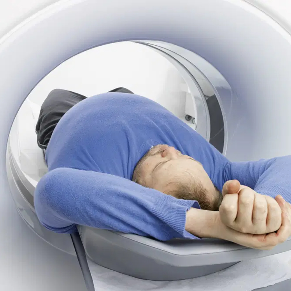
Patient Experience During QCT Scanning
During Quantitative Computed Tomography (QCT) scanning, patients typically experience a similar process to traditional CT scans. They are positioned on a table that moves through the CT scanner, which resembles a large ring or doughnut-shaped machine. The patient may need to hold their breath briefly during the scan to minimize motion artifacts and ensure image quality. The scanning process itself is painless and non-invasive, with the entire procedure usually lasting only a few minutes.
Patients may be required to remain still during the scan to obtain clear images, and they may hear humming or buzzing noises as the machine operates. Radiologic technologists or healthcare professionals performing the scan will communicate with the patient throughout the process, providing instructions and ensuring their comfort and safety.
Overall, the patient experience during QCT scanning is generally well-tolerated, with minimal discomfort. However, individuals with claustrophobia or anxiety may experience some apprehension, which can usually be addressed through communication with the healthcare team beforehand.
The Importance and Purpose of Bone Density Measurement
Bone density measurement holds significant importance in assessing bone health and diagnosing various conditions related to bone strength and integrity. The primary purpose of bone density measurement is to evaluate the density and strength of bones, particularly in the context of osteoporosis, a common bone disease characterized by low bone mass and deterioration of bone tissue.
Accurate measurement of bone density is essential for:
Early Detection of Osteoporosis: Bone density measurement helps in the early detection of osteoporosis, allowing for timely intervention and management to prevent fractures and other complications associated with weakened bones.
Assessment of Fracture Risk: Low bone density is a major risk factor for fractures, especially in the spine, hips, and wrists. Bone density measurement aids in assessing an individual’s risk of fracture and guiding preventive measures to reduce fracture incidence.
Monitoring Bone Health: Bone density measurement is used to monitor changes in bone density over time, providing valuable information about bone health and response to treatment interventions for conditions such as osteoporosis.
Guiding Treatment Decisions: Bone density measurement helps healthcare providers determine the most appropriate treatment strategies for individuals with osteoporosis or other bone-related conditions. Treatment options may include lifestyle modifications, medications to increase bone density, and fall prevention strategies.
Evaluation of Bone Health in High-Risk Populations: Bone density measurement is particularly important in high-risk populations, such as postmenopausal women, older adults, individuals with a family history of osteoporosis, and those with certain medical conditions or lifestyle factors that predispose them to bone loss.
Overall, bone density measurement plays a crucial role in the prevention, diagnosis, and management of bone-related conditions, contributing to improved patient outcomes and quality of life. By accurately assessing bone density, healthcare providers can identify individuals at risk of fractures, implement preventive measures, and tailor treatment interventions to optimize bone health.
Use of QCT in the Diagnosis of Osteoporosis and Other Bone Diseases
Quantitative Computed Tomography (QCT) is employed in diagnosing osteoporosis and other bone diseases by providing accurate measurements of bone density. QCT enables early detection of osteoporosis by precisely measuring bone mineral density (BMD) in key areas like the spine, hip, and wrist. It detects subtle changes in bone density, allowing for timely intervention before significant bone loss occurs. By assessing fracture risk based on BMD measurements, QCT aids clinicians in identifying individuals who may benefit from preventive measures to reduce fracture incidence. Additionally, QCT is valuable for monitoring disease progression, evaluating treatment effectiveness, and diagnosing various bone disorders beyond osteoporosis, such as osteopenia, metabolic bone diseases, and bone tumors. Overall, QCT’s precise assessment of bone density significantly enhances the diagnosis and management of osteoporosis and other bone-related conditions.
Quantitative Computed Tomography (QCT) is utilized for bone density measurement through a process that involves several steps:
Image Acquisition: During a QCT scan, the patient lies on a table that passes through a CT scanner. The scanner emits X-ray beams that penetrate the body and are attenuated differently as they pass through bone and soft tissues.
Attenuation Measurement: The CT scanner detects the attenuation of X-ray beams as they pass through the body. The attenuation values are measured in Hounsfield units (HU), which quantify the X-ray attenuation properties of different materials.
Calibration: To convert Hounsfield units into bone mineral density (BMD) measurements, the CT scanner is calibrated using standard phantoms with known densities of hydroxyapatite, a major component of bone.
BMD Calculation: The attenuation values obtained from the patient’s scan are compared to the attenuation values of the calibration phantoms. By correlating these values, the CT scanner calculates the BMD of the scanned bone tissue in units of milligrams per cubic centimeter (mg/cm³) or grams per cubic centimeter (g/cm³).
Analysis and Interpretation: The resulting BMD measurements provide quantitative information about the density and mineral content of the bone tissue. These measurements are analyzed by healthcare professionals to assess bone health, diagnose conditions such as osteoporosis, and evaluate fracture risk.
Overall, QCT enables accurate and precise measurement of bone density by quantifying the attenuation of X-ray beams passing through bone tissue. This quantitative approach provides valuable information for assessing bone health and diagnosing bone-related conditions.
Quantitative Computed Tomography (QCT) scanning is utilized across medical specialties for various clinical applications:
Quantitative Computed Tomography (QCT) scanning is utilized across medical specialties for various clinical applications:
Osteoporosis Diagnosis and Management: QCT scanning assesses bone mineral density (BMD) and aids in diagnosing osteoporosis. It helps monitor disease progression and evaluate treatment effectiveness.
Fracture Risk Assessment: QCT scanning measures bone density in critical areas like the spine and hip, aiding in assessing fracture risk and guiding preventive measures and treatment interventions.
Orthopedic Surgery Planning: QCT provides detailed bone structure imaging, assisting orthopedic surgeons in surgical planning and assessing bone quality. It aids in preoperative bone density assessment and identifying optimal surgical approaches.
Oncology Treatment Planning: QCT is used in radiation therapy planning for cancer treatment. It helps delineate tumor boundaries, assess bone involvement, and minimize radiation exposure to healthy tissues.
Metabolic Bone Disorders Evaluation: QCT assists in evaluating metabolic bone disorders such as Paget’s disease and hyperparathyroidism. It provides quantitative bone density measurements, aiding in disease diagnosis and management.
Trauma and Sports Medicine: QCT scanning evaluates traumatic injuries, assesses bone fractures, and guides treatment decisions in trauma and sports medicine. It aids in identifying bone abnormalities and assessing healing progress after injuries.
Research and Clinical Trials: QCT is employed in research studies and clinical trials investigating bone health, osteoporosis treatments, and other musculoskeletal conditions. It provides quantitative measurements for evaluating treatment efficacy and disease progression.
Overall, QCT scanning has diverse clinical applications in diagnosing, monitoring, and managing various bone-related conditions across medical specialties, contributing to improved patient care and outcomes.
Quantitative Computed Tomography (QCT) plays a crucial role in the evaluation of post-traumatic bone deformities and injuries. Here’s how QCT is utilized in this context:
Detailed Imaging: QCT provides detailed three-dimensional imaging of bone structures, allowing for comprehensive evaluation of post-traumatic deformities and injuries. It offers superior visualization compared to conventional X-ray imaging, enabling healthcare providers to assess the extent and severity of the trauma.
Accurate Assessment: QCT enables accurate assessment of bone density and morphology, which is essential for evaluating post-traumatic bone deformities such as fractures, dislocations, and malunions. It helps in identifying subtle deformities and assessing the alignment and stability of fractured bones.
Fracture Healing Monitoring: QCT is used to monitor the healing process of fractures by assessing changes in bone density and structure over time. It provides quantitative measurements that aid in evaluating the effectiveness of treatment interventions and determining the progress of bone healing.
Surgical Planning: QCT imaging assists orthopedic surgeons in surgical planning for the correction of post-traumatic bone deformities. It provides detailed information about the location, extent, and severity of deformities, guiding the selection of appropriate surgical techniques and approaches.
Treatment Evaluation: QCT is valuable for evaluating the outcomes of treatment interventions for post-traumatic bone deformities and injuries. It helps healthcare providers assess the effectiveness of surgical procedures, such as fracture reduction and fixation, and non-surgical treatments like immobilization and physical therapy.
Long-Term Follow-Up: QCT imaging allows for long-term follow-up of patients with post-traumatic bone deformities and injuries. It enables healthcare providers to monitor changes in bone density and structure over time, assess the stability of treated fractures, and detect any complications or recurrence of deformities.
Overall, QCT is an indispensable tool in the evaluation and management of post-traumatic bone deformities and injuries, providing detailed imaging and accurate assessment that guide treatment decisions and optimize patient outcomes.
Quantitative Computed Tomography (QCT) offers several advantages and benefits in the assessment of bone density and various clinical applications:
Accurate Measurement: QCT provides precise and quantitative measurements of bone mineral density (BMD), offering superior accuracy compared to conventional methods such as dual-energy X-ray absorptiometry (DXA). This accuracy is particularly beneficial in diagnosing osteoporosis and assessing fracture risk.
Differentiation of Bone Types: QCT can differentiate between cortical and trabecular bone, providing separate measurements of their densities. This capability is essential for evaluating bone health comprehensively, as changes in cortical and trabecular bone density may have different clinical implications.
Assessment of Volumetric Bone Density: Unlike DXA, which measures areal bone density, QCT provides measurements of volumetric bone density. This comprehensive assessment accounts for bone size and structure, offering a more accurate representation of bone strength and fracture risk.
Evaluation of Bone Quality: QCT evaluates not only bone density but also bone quality, including parameters such as trabecular architecture and cortical thickness. This holistic assessment of bone health provides valuable insights into overall bone strength and fracture resistance.
Localization of Bone Density Measurements: QCT allows for precise localization of bone density measurements in specific regions of interest, such as the spine, hip, and wrist. This targeted assessment enables clinicians to focus on areas most relevant to osteoporosis diagnosis and fracture risk assessment.
Monitoring of Treatment Efficacy: QCT is valuable for monitoring the effectiveness of osteoporosis treatments and interventions. It allows clinicians to track changes in bone density over time, assess treatment response, and adjust management strategies accordingly.
Application in Various Clinical Settings: QCT has diverse clinical applications beyond osteoporosis assessment, including orthopedic surgery planning, oncology treatment planning, evaluation of metabolic bone disorders, and research studies. Its versatility makes it a valuable tool across different medical specialties.
Patient Comfort and Convenience: QCT scans are relatively quick and non-invasive, requiring minimal patient preparation and positioning. This enhances patient comfort and compliance, making QCT a preferred imaging modality for bone density assessment.
Overall, the advantages and benefits of QCT, including its accuracy, comprehensive assessment of bone density and quality, localization of measurements, and diverse clinical applications, contribute to its utility in enhancing the diagnosis, management, and treatment monitoring of various bone-related conditions.
Quantitative Computed Tomography (QCT) offers several advantages over other bone density measurement methods such as Dual-Energy X-ray Absorptiometry (DXA) and Quantitative Ultrasound (QUS):
Volumetric Bone Density Measurement: QCT provides volumetric bone density measurements, whereas DXA measures areal bone density. Volumetric measurements account for bone size and structure, offering a more comprehensive assessment of bone strength and fracture risk.
Differentiation of Cortical and Trabecular Bone: QCT can differentiate between cortical and trabecular bone, providing separate measurements of their densities. This differentiation is essential for assessing bone health comprehensively, as changes in cortical and trabecular bone density may have different clinical implications.
Accurate Assessment in Obese or Fragile Patients: QCT is less affected by soft tissue artifacts and body composition variations, making it suitable for accurately measuring bone density in obese patients or individuals with fragile bones.
Ability to Evaluate Bone Quality: QCT evaluates not only bone density but also bone quality, including parameters such as trabecular architecture and cortical thickness. This holistic assessment provides valuable insights into overall bone strength and fracture resistance.
Localization of Bone Density Measurements: QCT allows for precise localization of bone density measurements in specific regions of interest, such as the spine, hip, and wrist. This targeted assessment enables clinicians to focus on areas most relevant to osteoporosis diagnosis and fracture risk assessment.
Enhanced Diagnostic Accuracy: QCT offers superior diagnostic accuracy compared to DXA and QUS, particularly in diagnosing osteoporosis and assessing fracture risk. Its ability to provide detailed three-dimensional imaging and quantitative measurements contributes to enhanced diagnostic confidence.
Application in Various Clinical Settings: QCT has diverse clinical applications beyond osteoporosis assessment, including orthopedic surgery planning, oncology treatment planning, evaluation of metabolic bone disorders, and research studies. Its versatility makes it a valuable tool across different medical specialties.
Overall, the advantages of QCT over other bone density measurement methods, including its accuracy, comprehensive assessment of bone density and quality, differentiation of cortical and trabecular bone, precise localization of measurements, and diverse clinical applications, contribute to its utility in enhancing the diagnosis, management, and treatment monitoring of various bone-related conditions.
Quantitative Computed Tomography (QCT) scanning is generally considered safe, but like any medical procedure involving radiation, it carries some inherent risks. Here are the risks and safety considerations associated with QCT scanning:
Radiation Exposure: QCT scanning involves exposure to ionizing radiation, although the radiation dose is typically lower compared to conventional CT scans. However, repeated exposure to radiation over time may increase the risk of developing cancer. Healthcare providers aim to minimize radiation exposure by using the lowest effective dose necessary to obtain diagnostic images.
Allergic Reactions: Some patients may have allergic reactions to the contrast agents used in QCT scans. These reactions can range from mild itching or rash to more severe reactions such as anaphylaxis. It’s essential for healthcare providers to screen patients for allergies and take appropriate precautions when administering contrast agents.
Pregnancy Risk: Pregnant women are generally advised to avoid exposure to ionizing radiation, including QCT scanning, particularly during the first trimester when fetal organs are developing. If a QCT scan is deemed necessary during pregnancy, healthcare providers will carefully weigh the benefits of the scan against the potential risks to the fetus and take appropriate precautions to minimize radiation exposure.
Renal Function Impairment: Contrast agents used in QCT scans can potentially affect kidney function, particularly in patients with pre-existing renal impairment. Healthcare providers will assess kidney function before administering contrast agents and may adjust the dosage or choose alternative imaging methods if necessary.
Contrast-Related Complications: In some cases, contrast agents used in QCT scans may cause complications such as contrast-induced nephropathy or allergic reactions. Healthcare providers will monitor patients closely for any adverse reactions and provide appropriate treatment if complications occur.
Discomfort and Claustrophobia: Some patients may experience discomfort or claustrophobia during QCT scanning, particularly if they are required to remain still inside the scanner for an extended period. Healthcare providers will take steps to minimize discomfort and anxiety, such as providing support and reassurance throughout the procedure.
Overall, while QCT scanning is generally safe, healthcare providers must carefully consider the risks and benefits for each patient and take appropriate precautions to ensure patient safety during the procedure.
Quantitative Computed Tomography (QCT) scanning involves exposure to ionizing radiation, which necessitates careful consideration of radiation exposure and implementation of safety measures to minimize potential risks. Here’s an overview of radiation exposure and safety measures associated with QCT scanning:
Quantitative Computed Tomography (QCT) scanning involves exposure to ionizing radiation, which necessitates careful consideration of radiation exposure and implementation of safety measures to minimize potential risks.
Radiation Exposure: QCT scans use X-ray technology to generate detailed images of the body’s internal structures. While the radiation dose associated with QCT scans is generally lower than conventional CT scans, it still exposes patients to ionizing radiation. The amount of radiation exposure during a QCT scan depends on various factors, including the scanning protocol, the area of the body being scanned, and the individual’s body size.
Radiation Safety Measures: Optimization of Imaging Protocols: Healthcare providers use optimized imaging protocols to minimize radiation exposure while ensuring diagnostic image quality. This involves adjusting scanning parameters such as tube voltage, tube current, and scan duration to achieve the lowest effective radiation dose. Use of Shielding: Lead shielding may be used to protect sensitive organs from radiation exposure during QCT scanning. For example, lead aprons or shields may be placed over the thyroid gland, breasts, or reproductive organs to reduce radiation dose to these areas. Patient-Specific Dose Reduction: Healthcare providers tailor the radiation dose based on the patient’s age, weight, and medical history to minimize unnecessary exposure while still obtaining diagnostic images. Limiting Repeat Scans: Healthcare providers avoid unnecessary repeat scans and perform imaging studies judiciously to reduce cumulative radiation exposure over time. Pregnancy Screening: Pregnant women are screened for pregnancy before undergoing QCT scans, and if pregnancy is confirmed, alternative imaging methods that do not involve ionizing radiation are considered whenever possible to minimize fetal radiation exposure. Radiation Monitoring: Radiology departments implement radiation monitoring programs to track and record radiation doses received by patients during imaging procedures. This helps ensure that radiation exposure remains within acceptable limits and allows for dose optimization over time.
Patient Education: Patients undergoing QCT scans are informed about the benefits and risks of radiation exposure and encouraged to ask questions and express any concerns they may have. Patient education also includes instructions on how to prepare for the scan and what to expect during the procedure.
Overall, radiation exposure during QCT scanning is carefully managed through optimization of imaging protocols, implementation of safety measures, and patient education, ensuring that the benefits of the procedure outweigh the potential risks.
Ensuring patient safety during Quantitative Computed Tomography (QCT) scanning involves implementing several measures to minimize potential risks and ensure a positive experience for the patient. Here are some key measures for patient safety during QCT scanning:
Patient Screening and Assessment: Before the QCT scan, patients undergo thorough screening and assessment to identify any potential contraindications or factors that may affect the safety of the procedure. This includes assessing the patient’s medical history, current health status, allergies, and any prior adverse reactions to contrast agents or imaging procedures.
Patient Education: Patients receive detailed information about the QCT scanning procedure, including its purpose, benefits, potential risks, and what to expect during the scan. Patient education also includes instructions on any necessary preparations, such as fasting or discontinuing specific medications before the procedure.
Informed Consent: Patients provide informed consent before undergoing QCT scanning, indicating their understanding of the procedure, its risks and benefits, and their willingness to proceed.
Radiation Safety Measures: Healthcare providers take measures to minimize radiation exposure during QCT scanning, including optimizing imaging protocols, using appropriate shielding to protect sensitive areas of the body, and employing patient-specific dose reduction techniques.
Pregnancy Screening: Female patients of childbearing age are screened for pregnancy before undergoing QCT scanning. If pregnancy is confirmed or suspected, alternative imaging methods that do not involve ionizing radiation may be considered to minimize fetal exposure.
Monitoring and Support: Throughout the scanning procedure, healthcare providers monitor the patient’s vital signs and well-being to ensure their safety and comfort. Patients are provided with appropriate support and reassurance to address any concerns or anxiety they may have during the scan.
Post-Scan Monitoring: After the QCT scan, patients may be monitored for any immediate adverse reactions or complications. Healthcare providers remain vigilant for signs of allergic reactions or contrast-related complications, providing prompt medical intervention if necessary.
Follow-Up Care: Patients receive appropriate follow-up care after the QCT scan, including post-procedure instructions, any necessary medication adjustments, and guidance on resuming normal activities.
By implementing these measures, healthcare providers ensure the safety and well-being of patients undergoing QCT scanning, minimizing risks and optimizing the overall patient experience.
The evaluation of Quantitative Computed Tomography (QCT) scan results involves a thorough analysis of the obtained images and quantitative measurements to assess bone density and detect any abnormalities. Here’s how QCT scan results are typically evaluated:
Visual Assessment: Radiologists and other healthcare professionals visually examine the QCT images to evaluate bone density, structure, and morphology. They assess the presence of any abnormalities such as fractures, deformities, or lesions.
Quantitative Analysis: QCT provides quantitative measurements of bone mineral density (BMD) in specific regions of interest, such as the spine, hip, or wrist. These measurements are compared to established reference values or age-matched norms to determine if the patient’s bone density is within normal ranges or if there is evidence of osteoporosis or other bone diseases.
Comparison with Previous Scans: If available, QCT scan results are compared to previous scans to monitor changes in bone density over time. This longitudinal assessment helps evaluate disease progression, treatment effectiveness, and the stability of bone health.
Fracture Risk Assessment: Based on the quantitative measurements of BMD, healthcare professionals assess the patient’s risk of fractures, particularly in critical areas such as the spine and hip. Lower bone density measurements indicate an increased risk of fractures and may prompt further evaluation and intervention.
Integration with Clinical Information: QCT scan results are integrated with the patient’s clinical history, risk factors for bone-related conditions, and other diagnostic findings to provide a comprehensive assessment of bone health. This integration helps guide treatment decisions and management strategies tailored to the individual patient’s needs.
Reporting: A detailed report of the QCT scan findings, including quantitative measurements and any observed abnormalities, is generated and communicated to the referring physician or healthcare provider. The report may also include recommendations for further evaluation or management based on the scan results.
Overall, the evaluation of QCT scan results involves a combination of visual assessment, quantitative analysis, comparison with previous scans, fracture risk assessment, integration with clinical information, and comprehensive reporting to guide clinical decision-making and optimize patient care.
Understanding and interpreting Quantitative Computed Tomography (QCT) scan results involves a comprehensive analysis of the obtained images and quantitative measurements. Here’s a guide to understanding and interpreting QCT scan results:
Visual Analysis: Radiologists visually examine the QCT images to assess bone density, structure, and morphology. They identify any abnormalities such as fractures, deformities, or lesions.
Quantitative Measurements: QCT provides quantitative measurements of bone mineral density (BMD) in specific regions of interest, such as the spine, hip, or wrist. These measurements are expressed in units such as milligrams per cubic centimeter (mg/cm³) or grams per cubic centimeter (g/cm³).
Reference Values: The quantitative measurements of BMD are compared to established reference values or age-matched norms to determine if the patient’s bone density falls within normal ranges or if there are indications of osteoporosis or other bone diseases.
Fracture Risk Assessment: Lower bone density measurements obtained from QCT scans may indicate an increased risk of fractures, particularly in critical areas such as the spine and hip. Healthcare professionals assess the patient’s fracture risk based on these measurements and other clinical factors.
Comparison with Previous Scans: If available, QCT scan results are compared to previous scans to monitor changes in bone density over time. This longitudinal assessment helps evaluate disease progression, treatment effectiveness, and the stability of bone health.
Clinical Integration: QCT scan results are integrated with the patient’s clinical history, risk factors for bone-related conditions, and other diagnostic findings to provide a comprehensive assessment of bone health. This integration helps guide treatment decisions and management strategies tailored to the individual patient’s needs.
Report Interpretation: A detailed report of the QCT scan findings, including quantitative measurements and any observed abnormalities, is generated and communicated to the referring physician or healthcare provider. The report may also include recommendations for further evaluation or management based on the scan results.
Understanding and interpreting QCT scan results require expertise in radiology and bone health assessment. Healthcare professionals, particularly radiologists and orthopedic specialists, play a crucial role in interpreting scan results accurately and providing appropriate clinical recommendations based on the findings.
The use of Quantitative Computed Tomography (QCT) results in treatment planning involves leveraging the obtained bone density measurements and imaging findings to guide the management of various bone-related conditions. Here’s how QCT results are utilized in treatment planning:
Osteoporosis Management: QCT provides accurate measurements of bone mineral density (BMD), which are used to diagnose osteoporosis and assess fracture risk. Based on QCT results, healthcare providers can determine the appropriate treatment strategies, including pharmacological interventions, lifestyle modifications, and fall prevention measures, tailored to the patient’s individual risk profile.
Fracture Risk Assessment: QCT measurements of BMD, particularly in critical areas such as the spine and hip, help assess the patient’s risk of fractures. Healthcare providers use this information to implement preventive measures, such as calcium and vitamin D supplementation, weight-bearing exercises, and medication adjustments, to reduce the risk of future fractures.
Orthopedic Surgery Planning: In orthopedic surgery, QCT imaging provides detailed three-dimensional visualization of bone structures and accurate assessment of bone density. Surgeons use QCT results to plan surgical procedures, such as fracture reduction, fixation, and joint replacement, optimizing surgical approaches and implant selection based on the patient’s bone quality and anatomy.
Radiation Therapy Planning: QCT scans are used in radiation therapy planning for cancer treatment, particularly in cases where bone involvement is present. QCT results help delineate tumor boundaries, assess bone density, and determine the optimal radiation dose and treatment fields, minimizing radiation exposure to healthy tissues while maximizing tumor control.
Evaluation of Treatment Effectiveness: QCT scans are valuable for monitoring the effectiveness of treatment interventions, such as pharmacological therapies or surgical procedures, in improving bone density and reducing fracture risk. Serial QCT measurements allow healthcare providers to track changes in bone density over time and adjust treatment strategies as needed to optimize patient outcomes.
Research and Clinical Trials: QCT results are utilized in research studies and clinical trials investigating bone health, osteoporosis treatments, and other musculoskeletal conditions. QCT measurements provide quantitative data for evaluating treatment efficacy, assessing disease progression, and informing future treatment approaches.
Overall, QCT results play a critical role in treatment planning across various medical specialties, facilitating personalized and evidence-based management strategies for patients with bone-related conditions, including osteoporosis, fractures, and bone tumors.
Quantitative Computed Tomography (QCT) has current applications across various medical fields and holds significant potential for future advancements. Here’s an overview of both current applications and future potential of QCT:
Current Applications:
Osteoporosis Diagnosis and Management: QCT is widely used for diagnosing osteoporosis and assessing fracture risk by measuring bone mineral density (BMD). It provides more accurate measurements compared to conventional methods like Dual-Energy X-ray Absorptiometry (DXA).
Orthopedic Surgery Planning: QCT offers detailed three-dimensional imaging of bone structures, aiding in orthopedic surgery planning. Surgeons utilize QCT scans for preoperative assessment of bone density, guiding surgical approaches, and optimizing implant selection.
Radiation Therapy Planning: QCT is employed in radiation therapy planning for cancer treatment, especially when bone involvement is present. It helps in delineating tumor boundaries, assessing bone density, and optimizing radiation dose delivery while minimizing damage to surrounding healthy tissues.
Metabolic Bone Disorders Evaluation: QCT assists in evaluating metabolic bone disorders such as Paget’s disease, hyperparathyroidism, and osteomalacia. It provides quantitative measurements of bone density and aids in disease diagnosis and management.
Research Studies: QCT is used in various research studies investigating bone health, osteoporosis treatments, and musculoskeletal conditions. It provides quantitative data for evaluating treatment efficacy, assessing disease progression, and informing future research directions.
Future Potential:
Advanced Imaging Techniques: Continued advancements in QCT technology are expected to enhance imaging resolution, allowing for more detailed visualization of bone microarchitecture and subtle changes in bone density. This could improve diagnostic accuracy and treatment planning for various bone-related conditions.
Personalized Medicine: QCT holds the potential for personalized medicine approaches by providing quantitative measurements tailored to individual patient characteristics. This could lead to more precise assessment of fracture risk and personalized treatment strategies based on patient-specific bone density data.
Integration with Artificial Intelligence (AI): Integration of QCT with AI algorithms has the potential to streamline image analysis, automate quantitative measurements, and improve diagnostic accuracy. AI-based tools could assist radiologists in interpreting QCT scans and identifying abnormalities more efficiently.
Expanded Clinical Applications: QCT may find expanded clinical applications beyond bone health assessment. There is growing interest in utilizing QCT for assessing cardiovascular health, lung function, and soft tissue characterization, among other potential applications.
Portable QCT Devices: Development of portable QCT devices could enable point-of-care bone density assessment in clinical settings outside of traditional radiology departments. This could improve accessibility to bone health evaluation, particularly in underserved areas.
In summary, Quantitative Computed Tomography (QCT) has established itself as a valuable tool in bone health assessment with current applications in osteoporosis diagnosis, orthopedic surgery planning, radiation therapy planning, metabolic bone disorders evaluation, and research studies. Looking ahead, ongoing technological advancements and research efforts hold promise for expanding the scope of QCT applications and enhancing its role in personalized medicine and clinical practice.
Patient rights and important considerations regarding Quantitative Computed Tomography (QCT) scanning encompass various aspects aimed at ensuring patient safety, informed decision-making, and respect for individual autonomy. Here are key patient rights and considerations regarding QCT scanning:
Informed Consent: Patients have the right to receive detailed information about the purpose, risks, benefits, and alternatives of the QCT scanning procedure. Healthcare providers must obtain informed consent from patients before proceeding with the scan, ensuring that patients understand the procedure and can make voluntary decisions about their healthcare.
Privacy and Confidentiality: Patients have the right to privacy and confidentiality regarding their medical information, including QCT scan results. Healthcare providers must adhere to strict confidentiality policies and protect patients’ personal health information in accordance with relevant privacy regulations, such as the Health Insurance Portability and Accountability Act (HIPAA) in the United States.
Patient Education: Patients have the right to receive education about QCT scanning, including its purpose, how the procedure is performed, potential risks, and any necessary preparations. Healthcare providers should provide clear and understandable information to empower patients to make informed decisions about their healthcare.
Respect for Dignity and Autonomy: Patients have the right to be treated with dignity, respect, and autonomy throughout the QCT scanning process. Healthcare providers should involve patients in decision-making, respect their preferences and cultural beliefs, and ensure that they are comfortable and informed throughout the procedure.
Access to Medical Records: Patients have the right to access their medical records, including QCT scan results, upon request. Healthcare providers should facilitate timely access to medical records and ensure that patients understand their results and can discuss them with their healthcare providers.
Safety and Quality of Care: Patients have the right to receive safe and high-quality care during QCT scanning. Healthcare providers should adhere to established safety protocols, minimize risks associated with radiation exposure, and ensure that equipment is properly maintained and calibrated.
Right to Refuse or Withdraw Consent: Patients have the right to refuse or withdraw consent for QCT scanning at any time, without fear of coercion or retaliation. Healthcare providers should respect patients’ autonomy and provide alternative options or address any concerns they may have about the procedure.
Clear Communication: Patients have the right to clear communication from healthcare providers regarding their QCT scan results, including explanations of findings, implications for their health, and recommendations for further evaluation or treatment.
Overall, patient rights and considerations regarding QCT scanning encompass informed consent, privacy, education, respect for dignity and autonomy, access to medical records, safety, and clear communication. Upholding these rights and considerations is essential for promoting patient-centered care and ensuring positive experiences for patients undergoing QCT scanning.
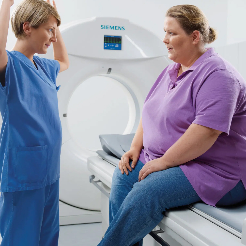
During the scanning process for Quantitative Computed Tomography (QCT), patient rights and preferences play a crucial role in ensuring a positive and comfortable experience. Here are some key patient rights and preferences to consider:
Informed Consent: Patients have the right to receive clear and understandable information about the QCT scanning procedure, including its purpose, benefits, potential risks, and alternatives. They should be given the opportunity to ask questions and provide informed consent before the procedure begins.
Privacy and Dignity: Patients have the right to privacy and dignity during the scanning process. Healthcare providers should ensure that patients are appropriately clothed and covered during the procedure to maintain their privacy. Additionally, patients should be provided with a private area to change clothes if necessary.
Comfort and Safety: Patients have the right to feel comfortable and safe during the scanning process. Healthcare providers should take measures to minimize any discomfort or anxiety experienced by patients, such as providing support pillows or blankets and explaining the procedure in advance to alleviate concerns.
Communication and Collaboration: Patients have the right to be actively involved in their care and decision-making during the scanning process. Healthcare providers should communicate openly and respectfully with patients, explaining each step of the procedure and addressing any concerns or preferences the patient may have.
Respect for Preferences: Patients have the right to express their preferences and concerns regarding the scanning process. Healthcare providers should respect patients’ preferences regarding factors such as the positioning during the scan, use of contrast agents, and any specific needs or accommodations they may require.
Access to Information: Patients have the right to access information about the scanning process, including details about the equipment used, potential sensations they may experience during the scan, and how long the procedure will take. This information can help alleviate anxiety and empower patients to feel more comfortable during the procedure.
Choice and Autonomy: Patients have the right to make choices about their care during the scanning process, including whether to proceed with the procedure, request breaks if needed, or ask questions about the procedure. Healthcare providers should respect patients’ autonomy and support their choices throughout the process.
Overall, respecting patient rights and preferences during the QCT scanning process is essential for promoting a positive patient experience and ensuring that patients feel informed, comfortable, and respected throughout the procedure.
Protecting and improving bone health is essential for overall well-being and longevity. Here are some key strategies and practices to protect and improve bone health:
Dietary Calcium and Vitamin D: Consuming an adequate amount of calcium and vitamin D is crucial for maintaining strong and healthy bones. Good dietary sources of calcium include dairy products, leafy greens, fortified foods, and certain types of fish. Vitamin D can be obtained through sunlight exposure and dietary sources such as fortified foods, fatty fish, and supplements if necessary.
Regular Exercise: Engaging in weight-bearing and resistance exercises helps strengthen bones and improve bone density. Weight-bearing exercises include walking, jogging, dancing, and stair climbing, while resistance exercises involve using weights or resistance bands to build muscle strength.
Maintaining a Healthy Weight: Maintaining a healthy weight through a balanced diet and regular physical activity is important for bone health. Excess body weight can increase the risk of osteoporosis and fractures, while being underweight may lead to bone loss and increased fracture risk.
Limiting Alcohol and Caffeine: Excessive alcohol consumption and high intake of caffeine can have negative effects on bone health. Limiting alcohol intake and moderating caffeine consumption from sources like coffee, tea, and energy drinks can help protect bone health.
Quitting Smoking: Smoking has been linked to decreased bone density and increased fracture risk. Quitting smoking can improve bone health and reduce the risk of osteoporosis and fractures.
Regular Bone Density Testing: Regular bone density testing, such as Dual-Energy X-ray Absorptiometry (DXA) or Quantitative Computed Tomography (QCT) scans, can help assess bone health and identify osteoporosis or osteopenia early. Based on the results, appropriate measures can be taken to prevent further bone loss and reduce fracture risk.
Medication Management: For individuals at high risk of osteoporosis or fractures, healthcare providers may recommend medications such as bisphosphonates, hormone therapy, or other bone-building medications. It’s important to follow healthcare provider recommendations and medication instructions carefully.
Fall Prevention: Taking steps to prevent falls can help protect bones from fractures, especially in older adults. This includes maintaining a safe home environment, using assistive devices if needed, wearing appropriate footwear, and participating in balance and strength training exercises.
Supplements and Medications: In some cases, supplements or medications may be recommended to support bone health, particularly for individuals with specific medical conditions or nutritional deficiencies. It’s important to consult with a healthcare provider before starting any new supplements or medications.
Regular Healthcare Visits: Regular check-ups with a healthcare provider can help monitor bone health and address any concerns or changes in bone density. Healthcare providers can provide personalized recommendations based on individual risk factors and health status.
By incorporating these strategies into daily life and working closely with healthcare providers, individuals can protect and improve their bone health, reducing the risk of osteoporosis and fractures and maintaining strong and healthy bones throughout life.
Based on Quantitative Computed Tomography (QCT) results, healthcare providers may recommend various interventions and measures to address specific findings and improve bone health. These recommendations are tailored to the individual patient’s needs and may include:
Lifestyle Modifications: Implementing lifestyle changes to support bone health, such as increasing calcium and vitamin D intake through diet or supplements, engaging in weight-bearing exercises, quitting smoking, limiting alcohol consumption, and taking precautions to prevent falls.
Pharmacological Interventions: Prescribing medications to improve bone density and reduce fracture risk in individuals diagnosed with osteoporosis or at high risk of fractures. These medications may include bisphosphonates, selective estrogen receptor modulators (SERMs), denosumab, or teriparatide, among others.
Fall Prevention Strategies: Recommending measures to prevent falls, such as removing hazards in the home environment, using assistive devices if necessary, wearing appropriate footwear, and participating in balance and strength training exercises.
Orthopedic Consultation: Referring patients to orthopedic specialists for further evaluation and management, especially in cases where QCT results indicate severe bone loss, structural abnormalities, or a high risk of fractures that may require surgical interventions.
Regular Monitoring: Scheduling follow-up appointments to monitor bone health and treatment effectiveness over time, which may include repeat QCT scans or other imaging studies to assess changes in bone density and fracture risk.
Education and Support: Providing patients with education about their QCT results, treatment options, and strategies for maintaining bone health. Offering support and guidance to help patients make informed decisions and adhere to recommended interventions.
These recommendations and measures are individualized based on the specific QCT findings and the patient’s overall health status, preferences, and risk factors for bone-related conditions. It’s essential for healthcare providers to work collaboratively with patients to develop personalized treatment plans that address their unique needs and goals for bone health.
Improving bone health involves adopting various measures and lifestyle changes aimed at maintaining strong and healthy bones. Here are some key measures to improve bone health:
Dietary Calcium and Vitamin D Intake: Consuming an adequate amount of calcium and vitamin D is essential for bone health. Good sources of calcium include dairy products (milk, cheese, yogurt), leafy green vegetables (kale, broccoli), fortified foods (orange juice, tofu), and certain types of fish (salmon, sardines). Vitamin D can be obtained through sunlight exposure and dietary sources such as fatty fish (salmon, mackerel), egg yolks, fortified foods (milk, cereal), and supplements if necessary.
Regular Physical Activity: Engaging in weight-bearing and muscle-strengthening exercises is crucial for building and maintaining bone density. Weight-bearing exercises include activities like walking, jogging, dancing, hiking, and stair climbing, while muscle-strengthening exercises involve resistance training using weights or resistance bands. Aim for at least 150 minutes of moderate-intensity aerobic activity or 75 minutes of vigorous-intensity aerobic activity per week, along with muscle-strengthening activities on two or more days per week.
Balanced Diet: Consuming a balanced diet rich in fruits, vegetables, whole grains, lean proteins, and healthy fats provides essential nutrients for bone health, including vitamins (C, K) and minerals (magnesium, phosphorus, potassium). Limiting excessive intake of sodium, processed foods, sugary beverages, and caffeine can also help preserve bone health.
Quit Smoking: Smoking has been linked to decreased bone density and an increased risk of fractures. Quitting smoking can improve bone health and reduce the risk of osteoporosis and fractures.
Limit Alcohol Consumption: Excessive alcohol consumption can negatively impact bone health by interfering with calcium absorption and increasing the risk of falls and fractures. Limit alcohol intake to moderate levels (up to one drink per day for women and up to two drinks per day for men).
Maintain a Healthy Weight: Maintaining a healthy weight through a balanced diet and regular physical activity is important for bone health. Being underweight can increase the risk of osteoporosis and fractures, while obesity can also affect bone health and increase the risk of other health conditions.
Ensure Safety: Take precautions to prevent falls and reduce the risk of fractures, especially in older adults. This includes maintaining a safe home environment by removing tripping hazards, using handrails and grab bars, wearing appropriate footwear, and participating in balance and strength training exercises.
Regular Bone Density Testing: For individuals at higher risk of osteoporosis or fractures, regular bone density testing (e.g., Dual-Energy X-ray Absorptiometry – DXA scans) can help assess bone health and guide preventive measures and treatment if necessary.
By incorporating these measures into daily life, individuals can improve bone health and reduce the risk of osteoporosis and fractures, leading to a healthier and more active lifestyle.
Frequently Asked Questions
Get a Free Second Opinion
Experienced Burtom Medical Team is Ready to Help
I consent to Burtom Health Group using my aforesaid personal data for the purposes described in this notice and understand that I can withdraw my consent at any time by sending a request to info@burtom.com.


