4D ultrasonography, also known as 4D ultrasound imaging or simply 4D ultrasound, is a medical imaging technique that provides a dynamic three-dimensional visualization of structures in real-time, allowing for the observation of motion within the body. It is an advanced form of traditional ultrasound that adds the dimension of time, allowing healthcare professionals to view moving images of internal organs and tissues.
This imaging modality works by emitting high-frequency sound waves into the body through a transducer. These sound waves bounce off internal structures and are then captured by the transducer, which converts them into electrical signals. The signals are processed by a computer to create real-time, three-dimensional images that can be viewed instantly on a monitor. The fourth dimension, time, is achieved by capturing a series of images over a short period, allowing the visualization of movement such as fetal development, cardiac function, or blood flow.
In summary, 4D ultrasonography enables healthcare providers to obtain detailed and dynamic images of anatomical structures and physiological processes, providing valuable information for diagnostic and therapeutic purposes in various medical fields, including obstetrics, gynecology, cardiology, and musculoskeletal imaging.
Basic Principles and Principles of 4D Ultrasonography: 4D ultrasonography operates on the foundational principles of traditional ultrasound imaging, with the added dimension of time. It utilizes high-frequency sound waves emitted from a transducer to penetrate body tissues, where they are reflected back as echoes. By precisely timing the emission and reception of these waves, along with sophisticated processing algorithms, 4D ultrasonography generates dynamic, real-time images that provide invaluable insights into the structure and function of internal organs and tissues.
Acquisition and Processing of Dynamic Images: The process of acquiring dynamic images in 4D ultrasonography involves emitting ultrasound waves into the body and capturing the reflected echoes. These echoes are then processed in real-time by advanced software algorithms, which assemble them into a sequence of images depicting the continuous movement and changes within the body over time. This real-time processing allows for the visualization of dynamic physiological processes such as fetal movement, cardiac function, and blood flow dynamics with remarkable detail and accuracy.
Applications and Importance of 4D Ultrasonography: 4D ultrasonography has diverse applications across various medical specialties, offering unparalleled insights into dynamic anatomical and physiological phenomena. In obstetrics, it plays a crucial role in monitoring fetal development and assessing maternal health, enabling early detection of abnormalities and ensuring optimal pregnancy outcomes. In cardiology, 4D ultrasonography facilitates the evaluation of cardiac function, myocardial perfusion, and valvular dynamics, aiding in the diagnosis and management of cardiovascular diseases. Additionally, it is employed in musculoskeletal imaging to visualize joint kinematics, soft tissue dynamics, and sports-related injuries, guiding treatment strategies and rehabilitation programs. Overall, 4D ultrasonography significantly enhances diagnostic capabilities, improves patient care, and contributes to advancements in medical research and clinical practice.
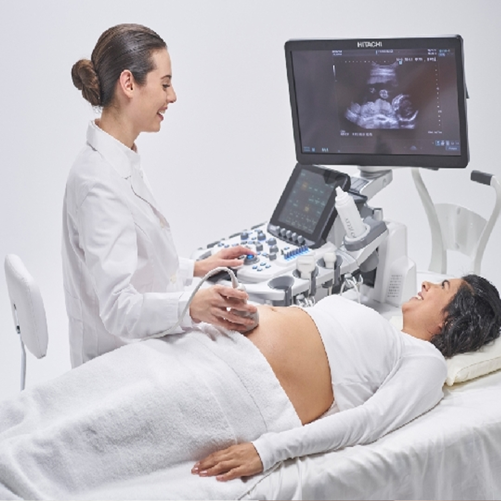
Monitoring Pregnancy and Fetal Development: 4D ultrasonography revolutionizes the way we monitor pregnancies and assess fetal development. By capturing dynamic, real-time images of the fetus in utero, it provides obstetricians and expecting parents with an immersive experience of the growing baby’s movements and behaviors. From the earliest stages of pregnancy to the final trimester, 4D ultrasonography enables detailed visualization of fetal anatomy, organ development, and physiological movements such as facial expressions, limb movements, and cardiac activity. This advanced imaging modality enhances prenatal care by facilitating early detection of potential abnormalities, guiding interventions, and fostering a deeper emotional connection between parents and their unborn child.
Technology and Working Principle of 4D Ultrasonography: The technology behind 4D ultrasonography builds upon the principles of traditional ultrasound imaging, incorporating the dimension of time to create dynamic, moving images. It relies on a transducer that emits high-frequency sound waves into the body, which then bounce off internal structures and tissues, producing echoes. These echoes are detected by the transducer and processed in real-time by sophisticated software algorithms. By precisely timing the emission and reception of ultrasound waves and analyzing the returning echoes, the system constructs a continuous stream of images that depict the dynamic movements and changes within the body over time. This real-time imaging capability provides invaluable insights into fetal development, allowing clinicians to monitor growth, assess organ function, and detect any anomalies early in pregnancy.
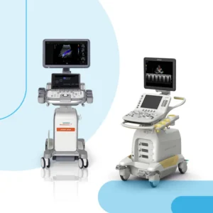
System and Basic Components of 4D Ultrasonography: A typical 4D ultrasonography system consists of several essential components designed to acquire, process, and display dynamic images of the fetus in utero. These components include the ultrasound transducer, which emits and receives ultrasound waves, the control panel, which allows the operator to adjust imaging parameters and settings, and the computer workstation, where the acquired images are processed and displayed in real-time. Additionally, specialized software algorithms are employed to enhance image quality, optimize visualization of fetal anatomy, and enable advanced imaging techniques such as volume rendering and multiplanar reconstruction. The integration of these components enables obstetricians to perform comprehensive fetal assessments, monitor pregnancy progression, and provide expecting parents with memorable visual experiences of their unborn child’s development.
Image Acquisition and Processing: 4D ultrasonography involves the acquisition and processing of dynamic images, providing real-time visualization of moving structures within the body. During image acquisition, the ultrasound transducer emits high-frequency sound waves into the body, which bounce off internal structures and tissues, generating echoes. These echoes are then detected by the transducer and processed in real-time by specialized software algorithms. The processed data is displayed as dynamic, moving images on a computer monitor, allowing clinicians to visualize anatomical structures and physiological movements in real-time.
Breast Cancer Screening and Diagnosis: In the field of breast imaging, 4D ultrasonography plays a crucial role in breast cancer screening and diagnosis. It offers high-resolution, real-time imaging of the breast tissue, allowing radiologists to detect and characterize suspicious lesions with precision. 4D ultrasonography is particularly valuable for evaluating breast masses, assessing their vascularity, and guiding image-guided biopsies for tissue sampling. Additionally, 4D ultrasonography can be used as a complementary imaging modality alongside mammography and magnetic resonance imaging (MRI) for comprehensive breast cancer screening and diagnosis.
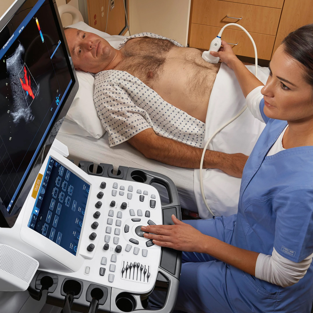
Cardiac and Vascular Examinations: In cardiac and vascular examinations, 4D ultrasonography provides valuable insights into cardiac anatomy, function, and blood flow dynamics. It enables clinicians to visualize the heart in motion, assessing chamber dimensions, wall motion abnormalities, and valvular function. Additionally, 4D ultrasonography is used in vascular examinations to evaluate blood vessels, detect abnormalities such as stenosis or aneurysms, and assess blood flow patterns. With its real-time imaging capabilities and high spatial resolution, 4D ultrasonography enhances the diagnosis and management of various cardiovascular conditions, including congenital heart defects, valvular disorders, and peripheral vascular diseases.
Real-Time and Dynamic Imaging: One of the primary advantages of 4D ultrasonography is its ability to provide real-time, dynamic imaging of internal structures and physiological movements within the body. Unlike traditional 2D ultrasound imaging, which captures static images at a single moment in time, 4D ultrasonography allows clinicians to visualize moving structures, such as fetal movements during pregnancy, cardiac motion, and blood flow dynamics. This real-time imaging capability enables clinicians to assess dynamic processes in real-time, enhancing diagnostic accuracy and enabling timely interventions.
Enhanced Visualization and Spatial Resolution: 4D ultrasonography offers enhanced visualization and spatial resolution compared to conventional 2D ultrasound imaging. The addition of the fourth dimension (time) allows for the reconstruction of volumetric datasets, providing a more comprehensive view of anatomical structures and physiological movements. With its high spatial resolution, 4D ultrasonography enables clinicians to visualize fine details and subtle abnormalities, improving diagnostic accuracy and confidence.
Non-Invasive and Radiation-Free: Another significant benefit of 4D ultrasonography is its non-invasive nature and lack of ionizing radiation exposure, making it safe for both patients and healthcare providers. Unlike other imaging modalities such as computed tomography (CT) or X-ray, which involve ionizing radiation, 4D ultrasonography uses harmless sound waves to generate images, posing no risk of radiation exposure. This makes 4D ultrasonography suitable for use in various patient populations, including pregnant women and pediatric patients, without concerns about radiation-related risks.
Improved Patient Experience: Due to its non-invasive nature and real-time imaging capabilities, 4D ultrasonography offers an improved patient experience compared to other imaging modalities. Patients undergoing 4D ultrasonography experience minimal discomfort during the procedure, as it does not involve injections, contrast agents, or ionizing radiation. Additionally, the ability to visualize moving structures in real-time provides patients with valuable insights into their condition, enhancing their understanding and engagement in the diagnostic process. Overall, the advantages of 4D ultrasonography contribute to its widespread use across various medical specialties for diagnostic imaging and monitoring purposes.
Radiation Exposure and Safety Measures: One of the notable advantages of 4D ultrasonography is its safety profile, as it does not involve ionizing radiation like some other imaging modalities such as X-ray or CT scans. Instead, 4D ultrasonography relies on harmless sound waves to generate images, eliminating the risk of radiation exposure for both patients and healthcare providers. This makes 4D ultrasonography a preferred imaging modality, especially for pregnant women and pediatric patients, as it poses no risk of radiation-related harm.
Precautions for Patient Safety During Scanning: While 4D ultrasonography is considered safe, healthcare providers should still take precautions to ensure patient safety during the scanning process. Proper positioning of the patient and the transducer is essential to obtain high-quality images while minimizing discomfort for the patient. Additionally, clinicians should adjust the ultrasound settings appropriately to avoid prolonged exposure to sound waves and minimize the risk of thermal effects, especially in sensitive areas such as the fetal brain during prenatal imaging. Regular monitoring of equipment performance and adherence to safety guidelines recommended by regulatory bodies further contribute to patient safety during 4D ultrasonography.
Evaluation and Interpretation of 4D Ultrasonography Images: Accurate interpretation of 4D ultrasonography images is crucial for making informed clinical decisions. Healthcare providers should undergo specialized training to acquire the necessary skills for image acquisition and interpretation. Proper orientation and labeling of anatomical structures, as well as recognition of normal variants and pathological findings, are essential for accurate diagnosis and patient management. Continuous quality assurance and peer review processes help ensure the reliability and consistency of image interpretation in 4D ultrasonography, enhancing diagnostic accuracy and patient care.
Understanding Image Results and Sharing with the Doctor: Understanding and interpreting 4D ultrasonography images require specialized knowledge and training. Clinicians must be able to identify anatomical structures and any abnormalities accurately. Collaborative efforts between radiologists, obstetricians, gynecologists, and other healthcare professionals ensure comprehensive evaluation and interpretation of images. Effective communication between the interpreting physician and the referring doctor is crucial for conveying diagnostic findings and formulating appropriate treatment plans.
Use and Monitoring of Images in the Treatment Process: 4D ultrasonography plays a vital role in guiding clinical decision-making and monitoring treatment response. Continuous imaging allows clinicians to assess dynamic changes in anatomical structures and monitor fetal growth and development during pregnancy. Additionally, real-time visualization of blood flow dynamics aids in evaluating cardiovascular function and diagnosing vascular abnormalities. Regular follow-up imaging sessions help track disease progression, evaluate treatment efficacy, and adjust therapeutic interventions as needed, contributing to improved patient outcomes.
Enhancements in Imaging Techniques and Future Potential: Advancements in imaging technology continue to enhance the capabilities of 4D ultrasonography. Ongoing research focuses on improving image resolution, enhancing tissue contrast, and developing advanced imaging modalities such as elastography and contrast-enhanced ultrasound. Integration of artificial intelligence (AI) algorithms and machine learning techniques holds promise for automated image analysis, pattern recognition, and decision support in clinical practice. These innovations have the potential to further enhance the diagnostic accuracy and clinical utility of 4D ultrasonography, paving the way for personalized medicine and precision healthcare delivery.
Patient Rights and Considerations Regarding 4D Ultrasonography: Respecting patient rights and ensuring their well-being are paramount in 4D ultrasonography practice. Patients have the right to receive clear and comprehensive information about the purpose, benefits, and potential risks of the procedure. Informed consent should be obtained before performing 4D ultrasonography, ensuring that patients understand the nature of the examination and any associated discomfort or limitations. Healthcare providers must prioritize patient comfort and privacy during imaging sessions, maintaining dignity and confidentiality throughout the process. Additionally, adherence to regulatory guidelines and ethical standards ensures that patient rights are upheld, promoting trust and transparency in healthcare delivery.

 4D ultrasound imaging, also known as four-dimensional ultrasound or real-time three-dimensional (3D) ultrasound, is a cutting-edge medical imaging technique that revolutionizes the visualization of anatomical structures. Unlike traditional 2D ultrasound, which provides static images, 4D ultrasound captures multiple consecutive 3D images in real-time, enabling dynamic visualization of moving structures. This innovative technology has significantly enhanced medical imaging capabilities, particularly in obstetrics and gynecology.