Burtom ‣ Technologies ‣ Digital Radiography (DR)
Language: 🇬🇧 English | 🇹🇷 Türkçe
Digital Radiography (DR) Overview

 Digital Radiography (DR) represents a revolutionary advancement in medical imaging technology, offering a sophisticated alternative to conventional film-based X-ray systems. Unlike traditional radiography, which relies on photographic films to capture and develop X-ray images, DR utilizes digital detectors that convert X-ray signals into digital data in real-time. This digital format enables immediate viewing and analysis of high-resolution images, eliminating the need for chemical processing and physical storage of films.
One of the key advantages of DR is its superior image quality and clarity. Digital detectors have a wider dynamic range and higher sensitivity compared to film, allowing for enhanced visualization of anatomical structures and abnormalities. This improved image quality facilitates more accurate diagnoses and treatment planning, leading to better patient outcomes.
In addition to superior image quality, DR offers several other benefits. The digital nature of DR allows for rapid image acquisition, reducing the time patients spend in imaging facilities and improving workflow efficiency for healthcare providers. Furthermore, DR systems can be integrated with picture archiving and communication systems (PACS), enabling seamless storage, retrieval, and sharing of images across healthcare networks. This facilitates collaboration among healthcare professionals and ensures timely access to patient imaging data for diagnostic and treatment purposes.
Another significant advantage of DR is its potential to lower radiation dose exposure for patients. Digital detectors are more sensitive to X-rays, requiring less radiation to produce high-quality images compared to traditional film-based systems. As a result, DR helps minimize radiation exposure while maintaining diagnostic image quality, contributing to patient safety and radiation protection efforts in healthcare settings.
Overall, Digital Radiography (DR) represents a transformative technology in diagnostic radiology, offering unparalleled image quality, efficiency, and patient safety compared to conventional film-based X-ray systems. Its widespread adoption across various healthcare settings underscores its significance in modern medical imaging and its crucial role in enhancing diagnostic accuracy and patient care.


What is Digital X-ray (DR) and How Does It Work?
Digital X-ray, also known as Digital Radiography (DR), is an advanced medical imaging technology used to produce high-quality X-ray images of the human body. Unlike traditional X-ray systems that use film to capture images, DR utilizes digital detectors to convert X-ray signals into digital images in real-time. This process involves passing X-rays through the body, which are absorbed differently by various tissues, creating variations in the intensity of the X-ray beam. These variations are then captured by the digital detectors, which convert them into digital data. This digital data is processed by the imaging system’s software to create detailed, high-resolution images that can be viewed instantly on a computer monitor. Digital X-ray systems offer several advantages over traditional film-based X-ray systems, including faster image acquisition, improved image quality, lower radiation dose exposure for patients, and the ability to digitally store, retrieve, and share images for immediate analysis by healthcare providers.
Basic Principles and Principles of Digital Radiography
Digital Radiography (DR) operates on the principle of using digital detectors to directly capture X-ray images of the human body in real-time, eliminating the need for film-based processing. This advanced imaging technique relies on the conversion of X-ray signals into digital data, which is processed by software to generate high-resolution images for diagnostic purposes.
Differences Between DR System and Conventional Radiography
The main differences between a Digital Radiography (DR) system and conventional radiography lie in the imaging technology and image processing methods. In a conventional radiography system, X-ray images are captured on film and require chemical processing, whereas DR systems use digital detectors to directly convert X-ray signals into digital images in real-time. This results in faster image acquisition, higher image quality, and the ability to digitally manipulate and enhance images for improved diagnostic accuracy. Additionally, DR systems offer lower radiation dose exposure for patients compared to conventional radiography.
Digital Radiography (DR) offers numerous advantages and benefits over conventional film-based radiography:
Faster Image Acquisition: DR systems provide immediate digital images, eliminating the need for film processing, which significantly reduces the time required to obtain diagnostic images.
Enhanced Image Quality: Digital detectors in DR systems offer higher sensitivity and dynamic range compared to film, resulting in sharper and more detailed images for improved diagnostic accuracy.
Lower Radiation Dose: DR systems require lower radiation doses to produce high-quality images compared to conventional radiography, reducing radiation exposure for patients and healthcare providers.
Image Manipulation and Enhancement: Digital images obtained through DR can be easily manipulated and enhanced using software tools, allowing for better visualization of anatomical structures and abnormalities.
Immediate Image Viewing and Analysis: DR images can be viewed instantly on computer monitors, enabling healthcare providers to quickly assess and diagnose medical conditions, leading to faster treatment decisions.
Digital Storage and Retrieval: DR images can be stored digitally in electronic health records (EHRs) and easily retrieved for future reference, facilitating efficient management of patient data and continuity of care.
Remote Image Sharing: Digital images obtained through DR can be easily shared with other healthcare providers for consultation and collaboration, regardless of geographical location, improving patient care coordination.
Overall, Digital Radiography offers significant advantages in terms of efficiency, image quality, radiation dose reduction, and data management, making it a preferred imaging modality in modern healthcare settings.
Faster and Easier Imaging
One of the key advantages of Digital Radiography (DR) is faster and easier imaging compared to conventional film-based radiography. With DR systems, X-ray images are captured digitally and instantly available for viewing on computer monitors, eliminating the need for film processing and reducing the time required to obtain diagnostic images. This rapid image acquisition streamlines the imaging process, allowing healthcare providers to quickly assess patient conditions and make timely treatment decisions, ultimately improving patient care and workflow efficiency in healthcare settings.
Image Enhancement and Digital Manipulation Capability
Digital Radiography (DR) offers the capability of image enhancement and digital manipulation, allowing for improved visualization and diagnostic accuracy. With DR systems, digital images can be easily manipulated using software tools to enhance contrast, adjust brightness and sharpness, and remove artifacts, thereby optimizing image quality for better visualization of anatomical structures and abnormalities. This digital manipulation capability enables radiologists and other healthcare providers to fine-tune images according to specific diagnostic needs, leading to more accurate diagnoses and better patient outcomes. Additionally, the ability to digitally manipulate images allows for precise measurements and annotations, facilitating communication and collaboration among healthcare professionals. Overall, the image enhancement and digital manipulation capability of DR systems contribute to improved diagnostic accuracy and efficiency in clinical practice.
Digital Radiography Technology and Working Principle
Digital Radiography (DR) technology operates on the principle of using digital detectors to directly capture X-ray images of the human body in real-time. Unlike conventional film-based radiography, where X-ray images are captured on film and require chemical processing, DR systems use digital detectors such as amorphous silicon or amorphous selenium to convert X-ray signals into digital data instantaneously.
The working principle of DR involves the following steps:
- X-ray Generation: X-rays are produced by an X-ray tube and directed towards the body part being imaged.
- X-ray Interaction: When X-rays pass through the body, they are attenuated differently by various tissues based on their density and composition.
- Digital Detection: Digital detectors, typically made of scintillator materials coupled to photodiodes or thin-film transistors, convert the attenuated X-ray signals into electrical charges.
- Signal Conversion: The electrical charges generated by the digital detectors are converted into digital data using analog-to-digital converters.
- Image Processing: The digital data is processed by computer algorithms to generate high-resolution X-ray images, which can be viewed instantly on computer monitors.
The digital nature of DR technology allows for immediate image acquisition, rapid data processing, and the ability to digitally manipulate and enhance images for improved diagnostic accuracy. Furthermore, DR systems offer advantages such as lower radiation dose exposure, faster imaging workflow, and seamless integration with electronic health records (EHRs) for efficient data management. Overall, Digital Radiography technology revolutionizes medical imaging by providing high-quality, real-time X-ray images with enhanced diagnostic capabilities.
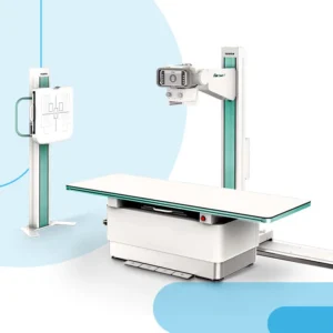
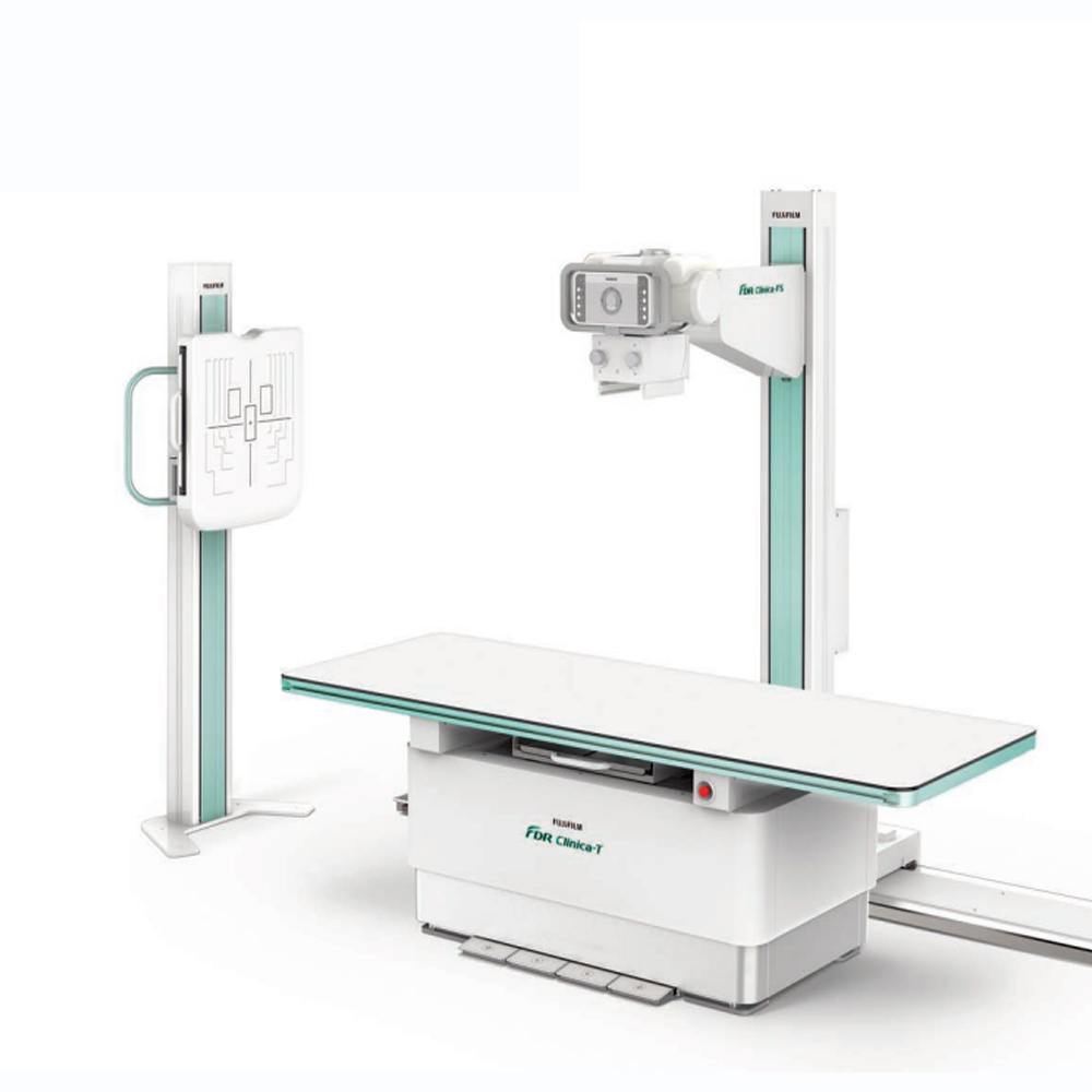
A Digital Radiography (DR) system consists of several basic components that work together to capture, process, and display X-ray images digitally. The main components of a DR system include:
X-ray Source: The X-ray source generates X-ray beams that pass through the body and interact with tissues to create images.
Detector Panel: The detector panel is a crucial component of a DR system, comprising digital detectors such as amorphous silicon or selenium. These detectors convert X-ray signals into digital data in real-time.
Image Acquisition System: The image acquisition system includes hardware and software components responsible for capturing and processing digital X-ray images. This system processes the digital data from the detector panel and converts it into high-resolution images.
Workstation: The workstation is a computer system equipped with specialized software for viewing, manipulating, and storing digital X-ray images. Radiologists and healthcare providers use the workstation to interpret images and make diagnostic decisions.
Display Monitor: The display monitor is an essential component of the DR system, where digital X-ray images are viewed by healthcare professionals. High-quality monitors with high resolution and brightness are used to ensure accurate interpretation of images.
Data Storage and Archiving: Digital X-ray images captured by the DR system are stored and archived in a Picture Archiving and Communication System (PACS) or other digital storage systems. This allows for easy access, retrieval, and management of patient images for future reference.
Control Console: The control console is a user interface that allows radiographers and technologists to control the X-ray source, adjust exposure parameters, and initiate image acquisition.
Overall, these basic components work together seamlessly to facilitate the efficient capture, processing, and interpretation of digital X-ray images in a Digital Radiography system.
Digital Radiography (DR) finds widespread clinical applications across various medical specialties due to its numerous advantages over conventional film-based radiography. Some common clinical applications and uses of DR include:
Diagnostic Imaging: DR is used for a wide range of diagnostic imaging procedures, including chest X-rays, skeletal imaging, abdominal imaging, and extremity imaging. It allows for high-quality, real-time imaging of internal structures to aid in the diagnosis of various medical conditions.
Trauma and Emergency Care: DR plays a critical role in trauma and emergency care settings by providing rapid imaging capabilities for assessing injuries such as fractures, dislocations, and soft tissue injuries. Its fast image acquisition time is especially valuable in time-sensitive situations.
Orthopedics: DR is extensively used in orthopedic imaging for evaluating bone fractures, joint injuries, and degenerative conditions such as arthritis. It enables detailed visualization of musculoskeletal structures to guide treatment planning and monitoring.
Dental Imaging: In dentistry, DR is used for dental radiography procedures such as intraoral and extraoral imaging, panoramic radiography, and cone-beam computed tomography (CBCT). It provides high-resolution images for diagnosing dental conditions and planning dental treatments.
Fluoroscopy: DR technology is employed in fluoroscopy procedures, allowing real-time visualization of moving anatomical structures such as the gastrointestinal tract, urinary tract, and cardiovascular system. It is used for procedures such as barium studies, voiding cystourethrography, and interventional radiology procedures.
Pediatric Imaging: DR is commonly used in pediatric imaging due to its lower radiation dose compared to conventional radiography. It enables detailed imaging of pediatric conditions such as growth plate injuries, congenital abnormalities, and respiratory conditions.
Screening and Preventive Care: DR is utilized in screening programs and preventive care initiatives for detecting conditions such as lung cancer, osteoporosis, and dental caries at an early stage. It enables healthcare providers to monitor changes in patient health over time and intervene proactively.
Overall, Digital Radiography has revolutionized medical imaging by offering enhanced imaging capabilities, faster image acquisition, lower radiation dose exposure, and improved workflow efficiency, making it indispensable in modern healthcare practice across various clinical specialties.
Digital Radiography (DR) is widely used for the diagnosis of injuries and fractures across various medical specialties. Some common applications include:
Trauma and Emergency Care: DR is an essential tool in trauma and emergency care settings for diagnosing fractures, dislocations, and soft tissue injuries resulting from accidents, falls, or sports-related injuries. It enables rapid imaging and immediate assessment of traumatic injuries, facilitating timely treatment decisions.
Orthopedics: DR plays a crucial role in orthopedic imaging for diagnosing fractures, evaluating joint injuries, and assessing degenerative conditions such as arthritis. It provides detailed images of bones and joints, allowing orthopedic specialists to determine the type and severity of fractures and plan appropriate treatment strategies, including surgical intervention if necessary.
Sports Medicine: In sports medicine, DR is used to diagnose musculoskeletal injuries, such as stress fractures, ligament tears, and muscle strains, commonly encountered in athletes. It helps sports medicine physicians and orthopedic specialists assess the extent of injuries, guide rehabilitation programs, and facilitate athletes’ return to sports activities safely.
Pediatrics: DR is commonly employed in pediatric imaging for diagnosing fractures and injuries in children and adolescents. It allows for accurate assessment of growth plate injuries, which are common in pediatric patients due to their developing skeletal system. Digital Radiography provides detailed images necessary for determining the appropriate management and follow-up of pediatric fractures.
Occupational Medicine: In occupational medicine, DR is used to evaluate work-related injuries, such as fractures caused by falls, accidents, or repetitive stress injuries. It aids occupational health professionals in diagnosing and managing workplace injuries, conducting pre-employment screenings, and assessing workers’ compensation claims.
Overall, Digital Radiography is a valuable diagnostic tool for the prompt and accurate diagnosis of injuries and fractures in various clinical settings, facilitating timely treatment and optimal patient care.
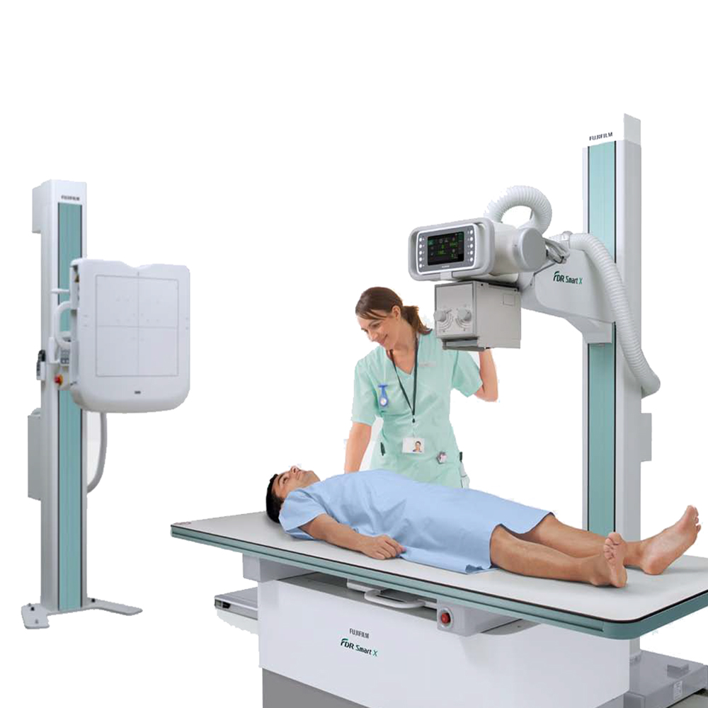
Patient preparation and experience during Digital Radiography (DR) scanning involve several steps to ensure optimal imaging quality and patient comfort. Here’s an overview:
Explanation: Before the procedure, the technologist explains the DR scanning process to the patient, including what to expect during the exam, the purpose of the exam, and any necessary preparations.
Clothing and Jewelry Removal: Patients may be asked to remove clothing, jewelry, and other metal objects that could interfere with the X-ray images. They are provided with a gown or drape to wear during the procedure.
Positioning: Patients are positioned by the technologist according to the specific imaging requirements for the area of the body being examined. Proper positioning is essential to obtain clear and accurate X-ray images.
Immobilization: In some cases, patients may need to remain still or be immobilized using positioning aids such as foam wedges or straps to minimize motion artifacts and ensure sharp images.
Shielding: Lead shields or aprons may be used to protect areas of the body not being imaged from unnecessary radiation exposure, particularly in sensitive areas such as the reproductive organs.
Communication: Throughout the procedure, the technologist communicates with the patient, providing instructions and reassurance to help alleviate any anxiety or discomfort.
Image Capture: The technologist operates the DR equipment to capture X-ray images of the targeted body part. Patients may be asked to hold their breath briefly during image acquisition to minimize motion blur.
Post-Scan Instructions: After the procedure, patients are provided with any necessary post-scan instructions, such as resuming normal activities or any restrictions related to the imaging procedure.
Overall, patient preparation and experience during DR scanning aim to ensure safety, comfort, and cooperation while obtaining high-quality X-ray images for accurate diagnosis and treatment planning. Effective communication between the technologist and the patient is essential to address any concerns and optimize the overall imaging experience.
The pre-scanning preparation process for Digital Radiography (DR) typically involves the following steps:
Appointment Scheduling: Patients schedule an appointment for their DR scan through the healthcare facility’s scheduling system. This ensures that the imaging facility is prepared for their visit and that there is sufficient time allocated for the procedure.
Patient Information Collection: Prior to the appointment, patients may be asked to provide relevant medical history, including any previous imaging studies, current medications, allergies, and other pertinent information. This helps the healthcare team assess the patient’s condition and tailor the imaging protocol accordingly.
Insurance Verification: Patients may need to provide insurance information to verify coverage for the DR scan. This ensures that there are no billing issues and helps the patient understand any out-of-pocket expenses.
Pre-Scan Instructions: Patients receive instructions from the healthcare facility regarding any specific preparations needed before the DR scan. This may include instructions to refrain from eating or drinking certain substances, fasting for a specified period, or discontinuing certain medications that may interfere with the imaging procedure.
Dress Code: Patients are typically instructed to wear comfortable, loose-fitting clothing without any metal objects or jewelry that may interfere with the X-ray imaging. In some cases, patients may be provided with a gown or drape to wear during the procedure.
Arrival and Registration: On the day of the appointment, patients arrive at the imaging facility and complete the registration process, which may involve providing identification, insurance information, and signing consent forms.
Pre-Scan Assessment: Before the DR scan, patients may undergo a brief pre-scan assessment by a technologist or nurse to confirm their identity, review medical history, and address any concerns or questions the patient may have.
Patient Comfort Measures: Healthcare providers may take measures to ensure patient comfort during the pre-scanning preparation process, such as providing a comfortable waiting area, addressing any anxiety or apprehension the patient may have, and explaining the procedure in detail.
Overall, the pre-scanning preparation process aims to ensure that patients are adequately prepared for their DR scan, both physically and emotionally, and that the imaging procedure proceeds smoothly and safely. Effective communication and collaboration between the healthcare team and the patient are essential to optimize the pre-scan preparation process.
Ensuring patient comfort and convenience during Digital Radiography (DR) scanning is essential to optimize the imaging experience and facilitate cooperation. Here are some strategies to enhance patient comfort and convenience during the scanning process:
Clear Communication: Healthcare providers should communicate clearly with the patient before, during, and after the procedure. They should explain the purpose of the DR scan, the steps involved, and what the patient can expect during the imaging process. Addressing any concerns or questions the patient may have can help alleviate anxiety and improve cooperation.
Comfortable Environment: Creating a comfortable and welcoming environment in the imaging facility can help put patients at ease. This includes providing comfortable seating in waiting areas, maintaining a clean and organized facility, and ensuring a calm and reassuring atmosphere.
Efficient Workflow: Streamlining the workflow and minimizing waiting times can contribute to patient convenience and satisfaction. Healthcare providers should strive to adhere to scheduled appointment times, minimize delays, and efficiently move patients through the imaging process.
Patient Positioning and Support: Proper patient positioning during the DR scan is crucial for obtaining high-quality images. Healthcare providers should ensure that patients are positioned comfortably and supported adequately to minimize discomfort and prevent movement artifacts.
Consideration of Patient Needs: Healthcare providers should be attentive to individual patient needs and preferences during the scanning process. This includes addressing any physical limitations, providing accommodations for patients with mobility issues, and offering support to patients who may require assistance.
Warmth and Empathy: Demonstrating warmth, empathy, and compassion towards patients can help create a positive and supportive environment during the imaging procedure. Healthcare providers should be attentive to patients’ emotional needs, offer reassurance, and respond promptly to any concerns or discomfort.
Monitoring Patient Comfort: Healthcare providers should continuously monitor patient comfort and well-being throughout the scanning process. They should encourage open communication and actively seek feedback from patients to address any discomfort or concerns promptly.
Overall, prioritizing patient comfort and convenience during Digital Radiography scanning involves creating a patient-centered approach that focuses on effective communication, a comfortable environment, efficient workflow, and individualized care. By implementing these strategies, healthcare providers can help ensure a positive and satisfactory imaging experience for patients undergoing DR scans.
The evaluation and interpretation of Digital Radiography (DR) images are critical steps in the diagnostic process, allowing healthcare providers to accurately assess the patient’s condition and formulate an appropriate treatment plan. Here’s an overview of the evaluation and interpretation process:
Image Quality Assessment: The first step in evaluating DR images is assessing their quality to ensure that they provide clear and accurate representations of the anatomical structures being imaged. Healthcare providers examine factors such as image clarity, resolution, contrast, and exposure to determine the overall quality of the images.
Anatomical Structures: Healthcare providers systematically review DR images to identify and assess the relevant anatomical structures, including bones, soft tissues, organs, and foreign objects. They evaluate the size, shape, alignment, and integrity of these structures for any abnormalities or pathology.
Pathological Findings: Healthcare providers analyze DR images to identify and characterize any pathological findings, such as fractures, dislocations, bone abnormalities, soft tissue injuries, foreign bodies, or signs of disease. They assess the location, extent, and severity of these findings to guide diagnosis and treatment decisions.
Comparison Studies: When available, healthcare providers may compare current DR images with previous imaging studies, such as X-rays or DR scans, to track changes over time and evaluate the progression of conditions or treatment outcomes. This comparative analysis provides valuable information for monitoring patient health and assessing treatment efficacy.
Radiological Reports: Based on their evaluation and interpretation of DR images, healthcare providers generate radiological reports summarizing their findings, observations, and diagnostic impressions. These reports provide detailed descriptions of any abnormalities or pathological findings identified on the images and may include recommendations for further evaluation or treatment.
Consultation and Collaboration: In complex cases or when additional expertise is needed, healthcare providers may consult with radiologists or other specialists to review DR images and obtain their expert opinion. Collaboration between healthcare providers ensures comprehensive evaluation and interpretation of DR images, leading to accurate diagnosis and appropriate management.
Overall, the evaluation and interpretation of DR images involve a systematic analysis of image quality, anatomical structures, pathological findings, and comparison studies to provide accurate diagnostic information and guide patient care effectively. Effective communication and collaboration among healthcare providers are essential for ensuring thorough evaluation and interpretation of DR images and delivering high-quality patient care.
Understanding Digital Radiography (DR) image results and effectively sharing them with the doctor are crucial steps in the diagnostic process, facilitating accurate diagnosis and treatment planning. Here’s how patients and healthcare providers can navigate this process:
Patient Understanding: Patients should actively engage in discussions with their healthcare providers to understand the results of their DR images. Healthcare providers should use layman’s terms and visual aids, such as annotated images or diagrams, to explain the findings in a clear and understandable manner. Patients should feel comfortable asking questions and seeking clarification about any aspects of their DR image results that they do not understand.
Interpretation by Healthcare Providers: Healthcare providers carefully review the DR images to identify any abnormalities or pathological findings. They interpret the images in the context of the patient’s medical history, symptoms, and clinical presentation. Healthcare providers use their expertise to formulate a diagnostic impression and treatment plan based on the DR image results.
Sharing with the Doctor: Healthcare providers communicate the findings of the DR images with the referring physician or specialist who ordered the imaging study. They provide a detailed radiological report summarizing the DR image results, including any abnormalities, pathological findings, and diagnostic impressions. The referring physician or specialist reviews the radiological report, discusses the findings with the patient, and incorporates the information into the overall management plan.
Collaboration and Follow-up: Effective communication and collaboration between healthcare providers, including radiologists, referring physicians, and specialists, are essential for ensuring comprehensive evaluation and management of the patient’s condition. Follow-up appointments may be scheduled to discuss treatment options, monitor progress, and adjust management plans based on the DR image results and clinical response.
Overall, patient understanding of DR image results and collaboration between healthcare providers are critical for delivering high-quality care and achieving optimal patient outcomes.
The utilization of Digital Radiography (DR) images is integral to the treatment process across various medical conditions. Here’s how these images contribute to treatment:
Diagnostic Insight: DR images aid in the accurate diagnosis of fractures, injuries, infections, tumors, and other abnormalities, providing clinicians with essential insights into the patient’s condition.
Tailored Treatment Plans: Based on DR image assessments, healthcare providers develop personalized treatment plans, considering the severity, location, and extent of pathological findings to determine the most suitable approach.
Treatment Progress Monitoring: Comparison of follow-up DR images with baseline or previous scans enables clinicians to monitor treatment efficacy, track condition progression, and make informed decisions regarding treatment adjustments.
Surgical Precision: In surgical settings, real-time DR images, like fluoroscopy, guide surgeons by visualizing anatomical structures, ensuring accurate instrument placement, monitoring progress, and enhancing surgical precision.
Patient Education: DR images serve as educational tools, allowing healthcare providers to illustrate pathological findings, explain treatment plans, and discuss potential outcomes with patients, facilitating informed decision-making.
DR images are indispensable in treatment planning, monitoring treatment effectiveness, guiding surgical interventions, and educating patients, ultimately contributing to enhanced patient outcomes and quality of care.
Digital Radiography (DR) is designed with several safety features to ensure patient well-being:
Enhanced Image Quality: DR systems allow for high-quality images to be produced using lower radiation doses compared to traditional film-based radiography.
Optimization of Exposure Parameters: Healthcare providers optimize exposure settings such as kilovoltage peak (kVp) and milliampere-seconds (mAs) to balance image quality with radiation dose, ensuring minimal exposure while maintaining diagnostic accuracy.
Automatic Exposure Control (AEC): DR equipment often includes AEC systems that adjust radiation dose based on patient anatomy, reducing unnecessary exposure and enhancing safety.
Compliance with Regulatory Standards: DR systems adhere to stringent regulatory standards set by organizations like the International Electrotechnical Commission (IEC) and the American Association of Physicists in Medicine (AAPM) to ensure safety and quality.
Quality Assurance Programs: Healthcare facilities implement quality assurance programs to regularly assess DR equipment performance, ensuring accurate imaging and patient safety.
Radiation Safety Training: Healthcare professionals operating DR equipment undergo specialized training in radiation safety and dose optimization, minimizing potential risks to patients.
Regular Equipment Maintenance: DR systems undergo regular maintenance and calibration to ensure optimal performance, reducing the likelihood of technical issues that could compromise patient safety.
Patient Education: Patients are informed about the benefits and risks of DR imaging, including radiation exposure, to enable them to make informed decisions about their healthcare.
By incorporating these safety measures and protocols, Digital Radiography ensures patient safety while providing accurate diagnostic imaging.
Ensuring patient safety during Digital Radiography (DR) scanning involves implementing various measures:
Positioning and Immobilization: Proper positioning and immobilization devices are used to ensure patients remain still during the procedure, reducing the likelihood of motion artifacts and the need for repeat scans.
Shielding: Lead shields or aprons are utilized to protect sensitive areas of the body from unnecessary radiation exposure, particularly in pediatric and pregnant patients.
Communication: Clear communication between healthcare providers and patients ensures that patients understand the procedure, follow instructions correctly, and feel comfortable throughout the scanning process.
Monitoring: Continuous monitoring of patients during the procedure allows healthcare providers to promptly address any discomfort or adverse reactions, ensuring patient safety and well-being.
Radiation Exposure Monitoring: Radiation exposure is carefully monitored and optimized to ensure that the lowest effective dose is used while maintaining image quality, thereby minimizing potential risks to patients.
Emergency Procedures: Healthcare facilities have established emergency procedures and protocols in place to address any unforeseen complications or medical emergencies that may arise during the scanning process.
Patient Education: Patients are educated about the procedure, including potential risks and safety measures, empowering them to actively participate in their healthcare decisions and advocate for their safety.
By implementing these measures, healthcare providers ensure patient safety and optimize the quality of Digital Radiography scans.
Patients undergoing Digital Radiography (DR) scanning have certain rights and should be informed about the procedure to ensure their safety and well-being. Here’s a summary of patient rights and essential information regarding DR scanning:
Informed Consent: Patients have the right to be informed about the purpose, risks, benefits, and alternatives of the DR scanning procedure before giving their consent.
Privacy and Confidentiality: Patients’ privacy and confidentiality must be respected throughout the DR scanning process. Healthcare providers should adhere to strict confidentiality protocols to protect patients’ personal health information.
Radiation Safety: Patients should be informed about the radiation exposure associated with DR scanning and any measures taken to minimize radiation dose while maintaining image quality.
Patient Comfort: Healthcare providers should prioritize patient comfort during the DR scanning procedure by ensuring proper positioning, providing clear instructions, and addressing any concerns or discomforts.
Right to Refuse: Patients have the right to refuse DR scanning if they have concerns or reservations about the procedure. Healthcare providers should respect patients’ decisions and provide alternative options when appropriate.
Access to Information: Patients have the right to access information about their DR scan results and discuss them with their healthcare provider. Clear communication and explanation of the results are essential for patients to understand their health status and any recommended follow-up actions.
Safety Measures: Patients should be informed about safety measures implemented during DR scanning, such as radiation shielding and monitoring, to ensure their safety and minimize potential risks.
By being informed about their rights and the DR scanning procedure, patients can actively participate in their healthcare decisions and contribute to a safe and positive imaging experience.
Privacy and data protection are critical considerations in Digital Radiography (DR) to safeguard patients’ personal health information and ensure compliance with privacy regulations. Here’s how privacy and data protection are addressed in DR:
Confidentiality: Healthcare providers must ensure the confidentiality of patients’ personal health information obtained during DR scanning. Access to patient data should be restricted to authorized personnel only, and measures such as password protection and encryption should be implemented to prevent unauthorized access.
Secure Data Storage: Patient data captured during DR scanning, including images and associated medical records, should be stored securely in electronic health record (EHR) systems or Picture Archiving and Communication Systems (PACS). These systems employ robust security measures to protect patient data from unauthorized access, loss, or theft.
Compliance with Regulations: Healthcare facilities must adhere to privacy regulations such as the Health Insurance Portability and Accountability Act (HIPAA) in the United States or the General Data Protection Regulation (GDPR) in the European Union. These regulations mandate strict guidelines for the collection, storage, and transmission of patient health information to ensure privacy and data protection.
Patient Consent: Patients should be informed about how their personal health information will be used and have the opportunity to provide consent for its collection, storage, and sharing. Consent forms should clearly outline the purposes of data collection and the measures in place to protect patient privacy.
Data Encryption: Patient data transmitted between DR systems and electronic health record systems should be encrypted to prevent interception and unauthorized access. Encryption algorithms ensure that patient information remains secure during transmission over networks.
Regular Audits and Monitoring: Healthcare facilities should conduct regular audits and monitoring of their data protection measures to identify and address any vulnerabilities or breaches. This proactive approach helps ensure ongoing compliance with privacy regulations and data protection standards.
By implementing robust privacy and data protection measures, healthcare providers can maintain patient trust, uphold confidentiality, and safeguard sensitive health information in Digital Radiography.
During the scanning process in Digital Radiography (DR), patients have certain rights and preferences that should be respected to ensure their comfort, safety, and overall well-being. Here are some key aspects of patient rights and preferences during DR scanning:
Informed Consent: Patients have the right to receive clear and comprehensive information about the DR scanning procedure, including its purpose, benefits, potential risks, and any alternatives. Informed consent should be obtained before proceeding with the scan, and patients should have the opportunity to ask questions and express any concerns.
Privacy and Dignity: Patients have the right to privacy and dignity during the scanning process. Healthcare providers should ensure that adequate measures are in place to maintain patient confidentiality and modesty throughout the procedure, including providing appropriate draping and minimizing exposure to other individuals.
Comfort and Communication: Patients should be made as comfortable as possible during the scanning process. Healthcare providers should communicate clearly with patients, explain the steps involved in the procedure, and address any anxieties or concerns that patients may have. Patients should also be encouraged to communicate their preferences and needs, such as requests for breaks or adjustments in positioning, to ensure a positive experience.
Safety and Well-being: Patients have the right to receive safe and high-quality care during DR scanning. Healthcare providers should adhere to strict safety protocols to minimize radiation exposure and ensure proper positioning to prevent injuries. Patients should be informed about safety measures in place, such as radiation shielding and monitoring, to reassure them of their safety.
Autonomy and Respect: Patients have the right to make autonomous decisions about their healthcare, including whether to undergo DR scanning. Healthcare providers should respect patients’ autonomy and preferences, including their right to refuse or request modifications to the procedure, as long as it does not compromise their safety or the quality of care provided.
Access to Information: Patients have the right to access information about their DR scan results and discuss them with their healthcare provider. Healthcare providers should provide patients with clear explanations of their results, address any questions or concerns, and involve them in decisions regarding further evaluation or treatment.
By respecting patients’ rights and preferences during the scanning process, healthcare providers can promote patient-centered care and enhance the overall experience for patients undergoing Digital Radiography.
The future of radiography techniques is poised for significant advancements driven by technological innovations and evolving healthcare needs. Some key developments shaping the future of radiography techniques include:
Artificial Intelligence (AI) Integration: AI-powered algorithms and machine learning techniques are increasingly being integrated into radiography systems to enhance image quality, improve diagnostic accuracy, and streamline workflow. AI-driven tools can assist radiologists in interpreting images, identifying abnormalities, and prioritizing cases, leading to more efficient and accurate diagnoses.
3D and Multi-dimensional Imaging: The adoption of three-dimensional (3D) and multi-dimensional imaging modalities, such as volumetric computed tomography (CT) and magnetic resonance imaging (MRI), is expanding the capabilities of radiography. These advanced imaging techniques offer enhanced visualization of anatomical structures, improved spatial resolution, and better characterization of pathology, enabling more precise diagnoses and treatment planning.
Portable and Point-of-Care Imaging: Portable radiography devices and point-of-care imaging solutions are becoming increasingly prevalent, allowing for bedside imaging in clinical settings such as intensive care units, emergency departments, and ambulatory care facilities. These portable systems offer real-time imaging capabilities, facilitating rapid diagnosis and treatment decisions, particularly in critical or emergency situations.
Digitalization and Connectivity: The ongoing digitalization of radiography systems and the integration of healthcare information technology (HIT) infrastructure are transforming the way radiographic images are acquired, stored, and shared. Digital radiography (DR) and picture archiving and communication systems (PACS) enable seamless image acquisition, remote access to images, and electronic sharing of patient data, improving collaboration among healthcare providers and enhancing patient care coordination.
Radiation Dose Reduction: Advances in radiography technology are focused on minimizing radiation dose exposure to patients and healthcare workers while maintaining diagnostic image quality. Techniques such as iterative reconstruction algorithms, dose modulation, and low-dose protocols are being implemented to optimize radiation dose levels without compromising diagnostic accuracy, thereby reducing the risk of radiation-related adverse effects.
Personalized Imaging and Precision Medicine: Radiography techniques are increasingly being tailored to individual patient characteristics and clinical needs, contributing to the advancement of precision medicine. Personalized imaging approaches, such as functional imaging and molecular imaging, enable the assessment of disease heterogeneity, treatment response monitoring, and the development of targeted therapeutic strategies tailored to each patient’s unique profile.
Overall, the future of radiography techniques is characterized by continued innovation, driven by advancements in technology, increasing demand for personalized healthcare solutions, and the evolving landscape of clinical practice. These developments hold the promise of enhancing diagnostic accuracy, improving patient outcomes, and transforming the delivery of radiology services in the years to come.
Advancements in Digital Radiography (DR) technology are continuously evolving to enhance imaging capabilities, improve workflow efficiency, and optimize patient care. Here are some key advancements and future directions in DR technology:
Direct Conversion Detectors: DR systems are increasingly utilizing direct conversion detectors, such as amorphous selenium (a-Se) or complementary metal-oxide-semiconductor (CMOS) sensors, to directly convert X-ray photons into electronic signals. These detectors offer higher sensitivity, improved image quality, and reduced electronic noise compared to indirect conversion detectors, leading to enhanced diagnostic accuracy.
Wireless and Portable DR Systems: The development of wireless and portable DR systems enables greater flexibility in imaging workflow, allowing for bedside imaging, point-of-care applications, and imaging in remote or challenging environments. These compact and lightweight systems offer real-time image acquisition, rapid image processing, and improved accessibility, particularly in emergency and critical care settings.
Dynamic Imaging Capabilities: DR technology is evolving to incorporate dynamic imaging capabilities, such as fluoroscopy and cine imaging, for real-time visualization of anatomical structures and physiological processes. Dynamic DR systems enable the assessment of moving structures, dynamic joint motion, and functional studies, providing valuable diagnostic information for musculoskeletal, cardiovascular, and gastrointestinal applications.
Artificial Intelligence (AI) Integration: AI algorithms and machine learning techniques are being integrated into DR systems to assist radiologists in image interpretation, workflow optimization, and decision support. AI-powered image analysis tools can aid in lesion detection, image reconstruction, artifact reduction, and automated image labeling, improving diagnostic efficiency and accuracy.
Low-Dose Imaging Protocols: DR technology is advancing to incorporate low-dose imaging protocols and dose optimization techniques to minimize radiation exposure to patients while maintaining diagnostic image quality. Innovations such as dose modulation, iterative reconstruction algorithms, and dose-tracking software enable radiation dose reduction without compromising diagnostic confidence, enhancing patient safety and radiation protection.
Enhanced Image Processing and Visualization: DR systems are incorporating advanced image processing algorithms and visualization tools to enhance image quality, improve soft tissue contrast, and reduce image artifacts. Features such as digital tomosynthesis, dual-energy imaging, and metal artifact reduction algorithms contribute to more detailed anatomical visualization and improved lesion detection.
Cloud-based Imaging Solutions: DR technology is shifting towards cloud-based imaging solutions, enabling seamless data storage, access, and sharing across healthcare facilities and remote locations. Cloud-based DR systems offer scalability, flexibility, and collaboration capabilities, facilitating remote interpretation, telemedicine consultations, and multidisciplinary care coordination.
Overall, advancements in DR technology are focused on improving imaging performance, enhancing workflow efficiency, and optimizing patient care through innovations in detector technology, imaging capabilities, AI integration, dose reduction techniques, and connectivity solutions. These advancements hold the promise of advancing diagnostic capabilities, improving clinical outcomes, and transforming the practice of digital radiography in the future.
Comparison with Other Imaging Techniques and Future Perspectives
Digital Radiography (DR) represents a significant advancement in medical imaging technology compared to conventional film-screen radiography and computed radiography (CR). It offers immediate image acquisition and digital storage, eliminating the need for film processing and handling, resulting in faster workflow and reduced turnaround time for patient diagnosis and treatment planning. DR systems utilize direct digital detectors, providing higher spatial resolution, improved contrast resolution, and better visualization of anatomical structures compared to CR systems. Additionally, DR offers greater flexibility in terms of portability, wireless connectivity, and point-of-care applications, making it ideal for use in various healthcare settings, including emergency departments, intensive care units, and outpatient clinics. Looking ahead, the future of DR technology is poised for further advancements, including improvements in detector technology, integration of artificial intelligence algorithms for image analysis, development of portable and handheld DR devices, implementation of low-dose imaging protocols, and enhanced connectivity with electronic health record systems and telemedicine platforms. These advancements are expected to enhance imaging capabilities, improve workflow efficiency, and optimize patient care in the field of radiology.
Frequently Asked Questions
Get a Free Second Opinion
Experienced Burtom Medical Team is Ready to Help
I consent to Burtom Health Group using my aforesaid personal data for the purposes described in this notice and understand that I can withdraw my consent at any time by sending a request to info@burtom.com.


