Burtom ‣ Technologies ‣ Dual-Energy X-ray Absorptiometry (DEXA)
Language: 🇬🇧 English | 🇹🇷 Türkçe
Dual-Energy X-ray Absorptiometry (DEXA) Overview

 Dual-Energy X-ray Absorptiometry (DEXA) is a sophisticated medical imaging technique employed for the evaluation of bone mineral density (BMD) and overall bone health. It operates by utilizing two X-ray beams with differing energy levels to discern between bone and soft tissue, allowing for accurate measurements of BMD in specific anatomical regions. DEXA scans are pivotal in the diagnosis and management of osteoporosis, a condition characterized by decreased BMD and increased susceptibility to fractures. By providing precise measurements of BMD, DEXA scans enable healthcare professionals to assess an individual’s risk of fractures and guide appropriate interventions to mitigate this risk.
Moreover, DEXA scans play a vital role in monitoring changes in bone density over time, allowing for the timely adjustment of treatment strategies to optimize bone health. Beyond osteoporosis, DEXA scans are also utilized in the evaluation of other bone-related conditions, such as osteopenia, hyperparathyroidism, and bone metastases. Additionally, DEXA scans are instrumental in assessing the efficacy of pharmacological interventions aimed at improving bone density and reducing fracture risk.
One of the key advantages of DEXA is its ability to provide precise and reliable measurements of BMD with minimal radiation exposure, making it a safe and widely-used imaging modality. The results obtained from DEXA scans assist healthcare providers in making informed decisions regarding the management of bone health and in implementing preventive measures to reduce the risk of fractures and associated complications. Overall, DEXA remains an indispensable tool in the field of bone health assessment, facilitating early diagnosis, risk stratification, and personalized management strategies for individuals at risk of bone-related disorders.


What is Bone Densitometry (DEXA) and How Does it Work?
Bone Densitometry, also known as Dual-Energy X-ray Absorptiometry (DEXA), is a medical imaging technique used to measure bone mineral density (BMD) and assess bone health. It operates by directing low-dose X-ray beams through the bones being examined. These beams are composed of two different energy levels, allowing for the differentiation between bone and soft tissue.
During the scan, the attenuation of each X-ray beam passing through the bone is measured. This attenuation is influenced by the bone mineral content and the thickness of the bone being examined. By comparing the attenuation of the two X-ray beams, DEXA can accurately calculate the bone mineral density of the scanned area.
DEXA scans are commonly used to diagnose osteoporosis, assess fracture risk, and monitor changes in bone density over time. They are particularly useful in evaluating the spine, hips, and wrists, which are common sites for osteoporotic fractures. DEXA scans are quick, non-invasive, and involve minimal radiation exposure, making them a safe and effective tool for evaluating bone health.
Basic Principles and Principles of DEXA
The basic principles and principles of Dual-Energy X-ray Absorptiometry (DEXA) revolve around its ability to accurately measure bone mineral density (BMD) and assess bone health. DEXA utilizes two X-ray beams with different energy levels to differentiate between bone and soft tissue. These beams pass through the bones being examined, and the attenuation of each beam is measured. By comparing the attenuation of the two X-ray beams, DEXA can calculate the bone mineral density of the scanned area.
DEXA operates on the principle that bone mineral content and bone thickness influence the attenuation of X-ray beams passing through the bone. The denser the bone, the greater the attenuation of the X-ray beams. This allows DEXA to provide precise and reliable measurements of BMD in specific regions of the body, such as the spine, hips, and wrists.
DEXA is widely used in clinical practice for the diagnosis of osteoporosis, assessment of fracture risk, and monitoring changes in bone density over time. It is considered the gold standard for BMD measurement due to its accuracy, precision, and ability to detect small changes in bone density.
Overall, the principles of DEXA revolve around its ability to accurately measure BMD by utilizing two X-ray beams with different energy levels and comparing their attenuation as they pass through bone tissue. This allows for the assessment of bone health and the early detection of conditions such as osteoporosis.
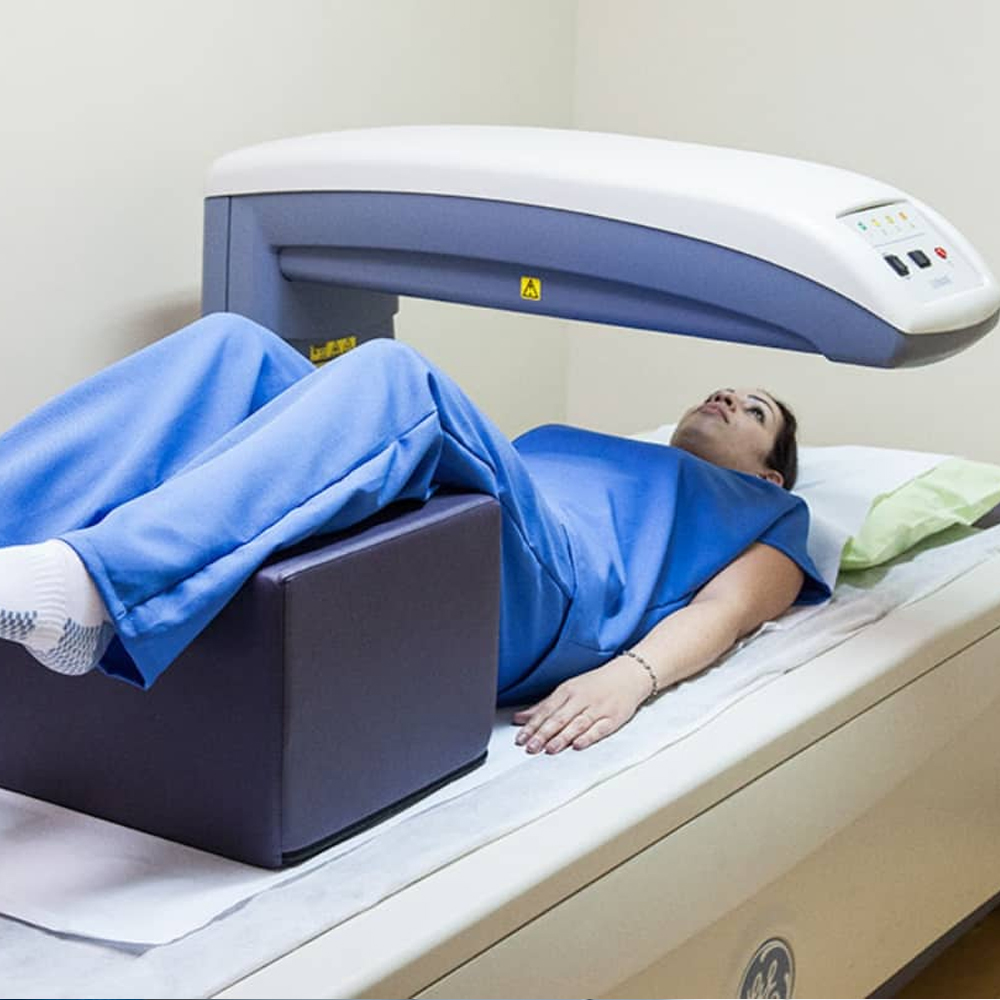
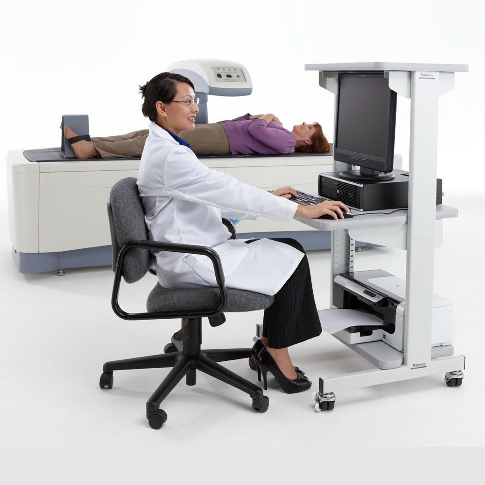
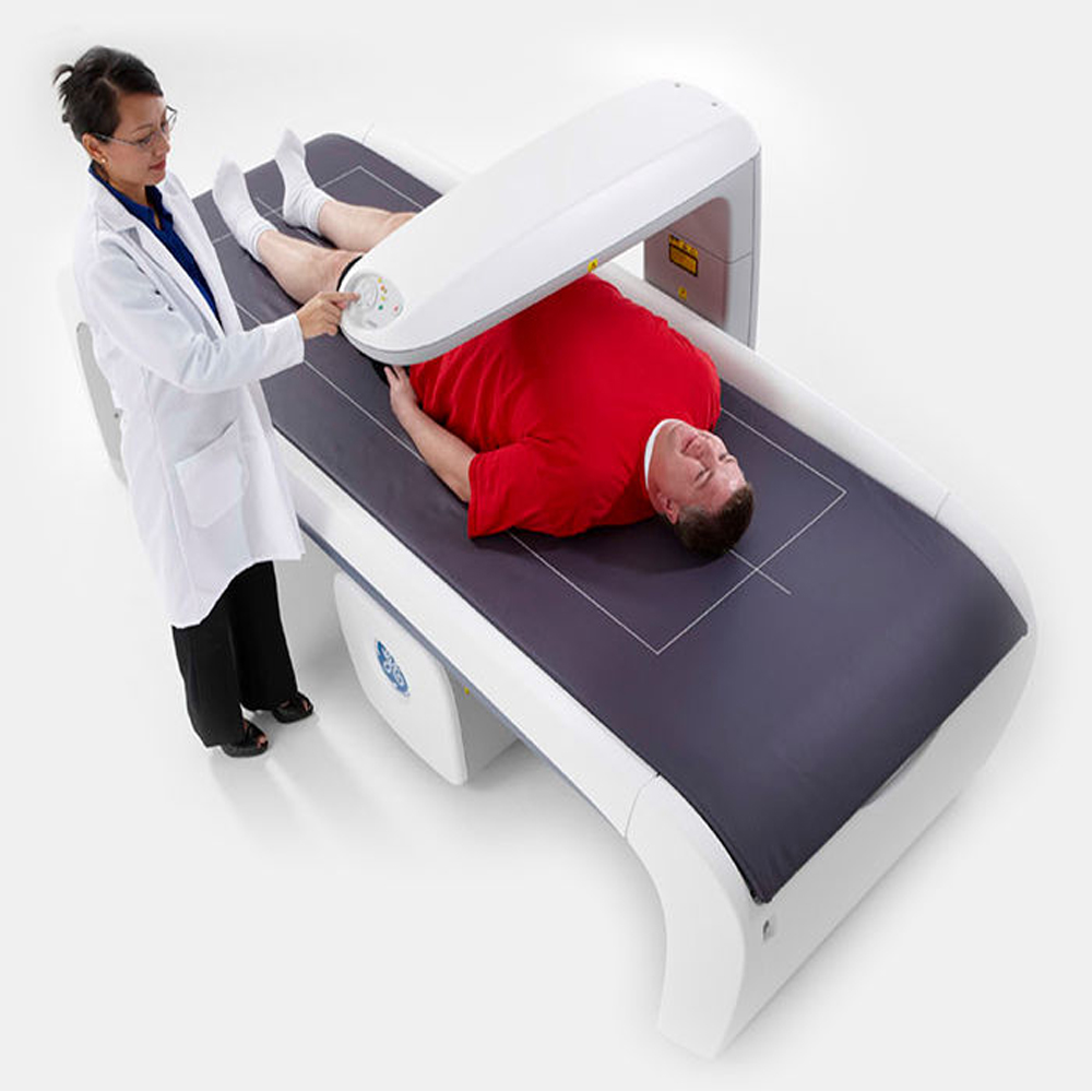
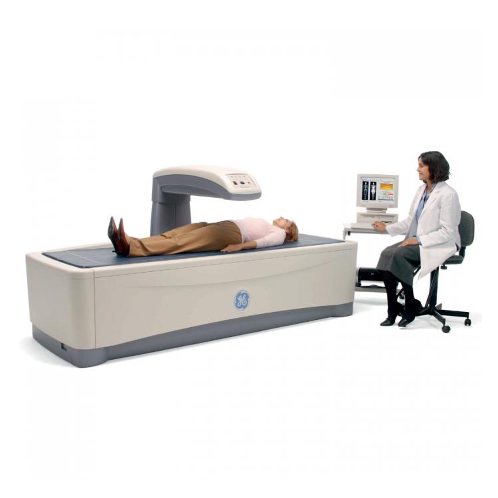
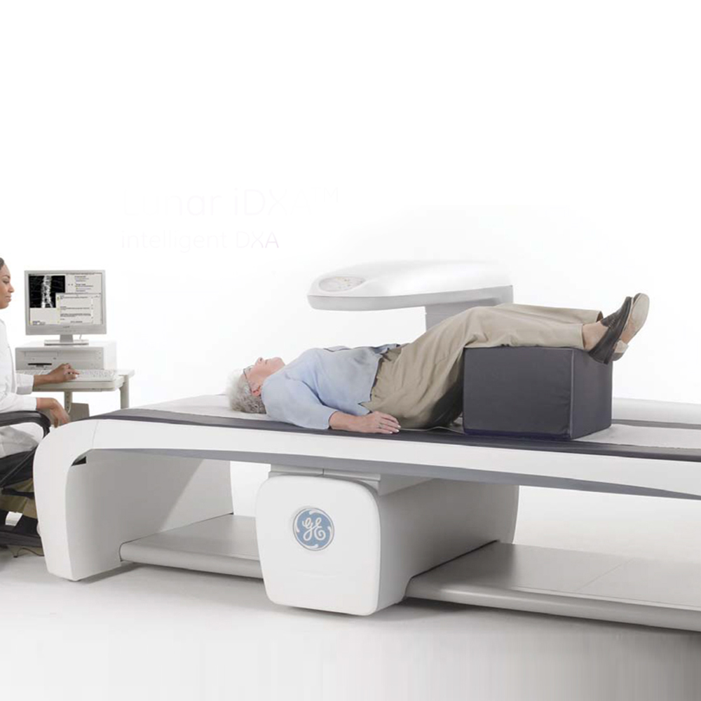
Patient Experience During DEXA Scanning
The patient experience during Dual-Energy X-ray Absorptiometry (DEXA) scanning is generally straightforward and well-tolerated. DEXA scanning is a quick and non-invasive procedure that typically lasts between 10 to 30 minutes, depending on the areas being scanned.
During the scan, the patient lies flat on a padded table while a mechanical arm containing the X-ray source and detector moves slowly over the body. The patient may be asked to remain still and hold their breath briefly to minimize movement and ensure clear images.
Patients may feel a slight pressure or sensation as the mechanical arm moves over their body, but the procedure is generally painless. Unlike other imaging techniques, such as MRI or CT scans, DEXA scanning does not involve any loud noises or confined spaces, contributing to a more comfortable experience for patients.
Additionally, DEXA scanning involves minimal radiation exposure, making it a safe imaging option for patients. After the scan is complete, patients can typically resume their normal activities immediately without any restrictions or recovery time.
Overall, the patient experience during DEXA scanning is typically comfortable, quick, and well-tolerated, with minimal discomfort or inconvenience. The procedure allows for accurate assessment of bone mineral density and plays a crucial role in the diagnosis and management of osteoporosis and other bone-related conditions.
Bone density measurement plays a crucial role in assessing bone health and diagnosing conditions such as osteoporosis. The importance and purpose of bone density measurement are multifaceted:
- Early Detection of Osteoporosis: Bone density measurement is essential for the early detection of osteoporosis, a condition characterized by low bone density and increased risk of fractures. Detecting osteoporosis early allows for timely intervention and management to prevent fractures and minimize complications.
- Fracture Risk Assessment: Bone density measurement helps assess an individual’s risk of fractures, particularly in weight-bearing bones such as the spine, hips, and wrists. Low bone density is associated with an increased risk of fractures, and accurate measurement helps identify individuals who may benefit from preventive measures and interventions to reduce fracture risk.
- Monitoring Bone Health: Bone density measurement is used to monitor changes in bone density over time, providing valuable information about the progression of bone loss and the effectiveness of treatment interventions. Regular monitoring helps healthcare providers assess treatment efficacy, adjust management strategies, and optimize bone health outcomes.
- Guiding Treatment Decisions: Bone density measurement guides treatment decisions in individuals at risk of osteoporosis or fracture. It helps healthcare providers determine the most appropriate management strategies, including lifestyle modifications, pharmacological interventions, and preventive measures to improve bone health and reduce fracture risk.
- Assessing Treatment Response: Bone density measurement is used to assess the response to treatment interventions aimed at improving bone health and reducing fracture risk. Monitoring changes in bone density over time helps evaluate treatment efficacy and guide adjustments in management strategies as needed.
- Risk Stratification and Prevention: Bone density measurement allows for risk stratification based on an individual’s fracture risk profile. Identifying individuals at higher risk of fractures enables targeted preventive measures, such as fall prevention strategies, exercise programs, and dietary modifications, to reduce the likelihood of fractures and improve overall bone health.
Overall, bone density measurement is essential for assessing bone health, diagnosing osteoporosis, evaluating fracture risk, guiding treatment decisions, monitoring treatment response, and implementing preventive measures to improve bone health outcomes and reduce the burden of fractures and associated complications.
Dual-Energy X-ray Absorptiometry (DEXA) plays a pivotal role in assessing bone health by providing accurate measurements of bone mineral density (BMD) in specific regions of the body. As a widely-used imaging technique, DEXA serves several important functions in the evaluation of bone health:
- Diagnosis of Osteoporosis: DEXA is the gold standard for diagnosing osteoporosis by measuring BMD and assessing bone quality. It helps identify individuals with low bone density who are at increased risk of fractures, enabling early intervention and treatment.
- Assessment of Fracture Risk: DEXA assists in assessing fracture risk by measuring BMD in key areas prone to fractures, such as the spine, hips, and wrists. Low BMD is associated with a higher risk of fractures, and DEXA helps identify individuals who may benefit from preventive measures to reduce fracture risk.
- Monitoring Changes in Bone Density: DEXA is used to monitor changes in BMD over time, providing valuable information about bone loss or improvement. Regular DEXA scans help track the progression of osteoporosis and evaluate the effectiveness of treatment interventions in maintaining or improving bone health.
- Evaluation of Treatment Response: DEXA plays a crucial role in evaluating the response to treatment interventions aimed at improving bone health. By measuring changes in BMD after treatment, DEXA helps healthcare providers assess treatment efficacy and make informed decisions regarding ongoing management strategies.
- Risk Stratification and Prevention: DEXA assists in risk stratification based on BMD measurements, helping identify individuals at higher risk of fractures. This enables targeted preventive measures, such as lifestyle modifications, exercise programs, and pharmacological interventions, to reduce fracture risk and improve overall bone health.
- Research and Clinical Trials: DEXA is widely used in research studies and clinical trials focused on bone health, osteoporosis, and related conditions. It provides precise and reliable measurements of BMD, contributing to advancements in the understanding and management of bone-related disorders.
Overall, DEXA plays a critical role in assessing bone health by providing accurate measurements of BMD, evaluating fracture risk, monitoring changes in bone density over time, assessing treatment response, enabling risk stratification, and contributing to research efforts aimed at improving bone health outcomes.
Determining osteoporosis and fracture risk is of paramount importance in healthcare, and Dual-Energy X-ray Absorptiometry (DEXA) plays a crucial role in this process. Osteoporosis is a silent disease characterized by low bone density and deterioration of bone tissue, leading to an increased risk of fractures, particularly in the spine, hips, and wrists. Here’s why determining osteoporosis and fracture risk is essential:
- Early Detection: Identifying osteoporosis early allows for timely intervention and management to prevent fractures and minimize complications. DEXA enables healthcare providers to accurately diagnose osteoporosis by measuring bone mineral density (BMD) and assessing fracture risk.
- Fracture Prevention: Osteoporotic fractures are associated with significant morbidity, mortality, and healthcare costs. Determining fracture risk through DEXA helps healthcare providers implement preventive measures, such as lifestyle modifications, fall prevention strategies, and pharmacological interventions, to reduce the likelihood of fractures and their associated complications.
- Treatment Planning: Knowing a patient’s fracture risk based on DEXA results guides treatment planning and management decisions. Individuals at higher risk of fractures may benefit from more aggressive treatment interventions, including pharmacological therapies aimed at improving bone density and reducing fracture risk.
- Monitoring Bone Health: DEXA is used to monitor changes in BMD over time, allowing healthcare providers to track the progression of osteoporosis and evaluate the effectiveness of treatment interventions. Regular monitoring helps optimize management strategies and maintain or improve bone health.
- Personalized Medicine: Determining osteoporosis and fracture risk through DEXA enables personalized medicine approaches tailored to individual patient needs. Healthcare providers can assess each patient’s risk profile and develop targeted interventions to reduce fracture risk and improve overall bone health outcomes.
- Quality of Life: Osteoporotic fractures significantly impact quality of life, causing pain, disability, loss of independence, and decreased mobility. By identifying individuals at risk of fractures through DEXA, healthcare providers can implement measures to preserve bone health, reduce fracture risk, and enhance quality of life.
In summary, determining osteoporosis and fracture risk is essential for early intervention, fracture prevention, treatment planning, personalized medicine, and improving quality of life. DEXA plays a critical role in accurately assessing bone health and guiding clinical decision-making to optimize patient outcomes.
DEXA (Dual-Energy X-ray Absorptiometry) technology operates on the principle of X-ray attenuation to measure bone mineral density (BMD). The working principle involves the following components and processes:
- X-ray Source and Detector: DEXA systems consist of an X-ray source that emits two different energy levels of X-ray beams and a detector that measures the attenuation of these beams after passing through the body.
- Calculation of Attenuation: As the X-ray beams pass through the body, they are attenuated differently by bone and soft tissue due to their varying densities. The detector measures the amount of X-ray energy that is absorbed by the bone tissue and soft tissue.
- Differentiation of Bone and Soft Tissue: By comparing the attenuation of the two X-ray beams, DEXA can differentiate between bone and soft tissue. This allows for the precise measurement of bone mineral content in specific regions of interest, such as the spine, hips, and wrists.
- Calculation of Bone Mineral Density: DEXA calculates bone mineral density (BMD) based on the attenuation of the X-ray beams passing through the bone tissue. BMD is expressed in grams per square centimeter (g/cm²) or as a T-score, which compares an individual’s BMD to that of a young, healthy adult of the same sex and ethnicity.
- Image Reconstruction and Analysis: DEXA systems reconstruct images of the scanned area based on the attenuation measurements. These images provide visual representations of bone density distribution and allow healthcare providers to analyze BMD measurements and identify regions of low bone density or osteoporosis.
- Software Algorithms: DEXA systems utilize specialized software algorithms to process the attenuation data, calculate BMD values, and generate comprehensive reports. These reports provide quantitative measurements of BMD, including T-scores and Z-scores, which are used for diagnosis and clinical interpretation.
Overall, DEXA technology utilizes the principle of X-ray attenuation to accurately measure bone mineral density in specific regions of the body. By differentiating between bone and soft tissue and calculating BMD values, DEXA plays a crucial role in the assessment of bone health and the diagnosis of conditions such as osteoporosis.
A DEXA (Dual-Energy X-ray Absorptiometry) system consists of several key components essential for accurate bone density measurement. These components work together to produce detailed images and quantitative measurements of bone mineral density (BMD). The basic components of a DEXA system include:
- X-ray Source: The X-ray source emits two distinct energy levels of X-ray beams that pass through the body during the scan. These X-ray beams are essential for differentiating between bone and soft tissue based on their attenuation properties.
- Detector: The detector, positioned opposite the X-ray source, measures the attenuation of the X-ray beams after they pass through the body. It captures the intensity of the X-ray beams and converts it into electrical signals that are processed by the system’s computer.
- Patient Table: The patient table is where the individual undergoing the DEXA scan lies during the procedure. It is typically padded and adjustable to ensure the patient’s comfort and proper positioning for accurate imaging.
- Mechanical Arm: The mechanical arm houses the X-ray source and detector and is responsible for their movement during the scan. It moves smoothly over the patient’s body, emitting X-ray beams and capturing attenuation data at various locations.
- Control Console: The control console serves as the interface for operating the DEXA system. It allows the technologist to control the scanning parameters, adjust settings, and monitor the scan in real-time.
- Computer System and Software: The computer system processes the attenuation data captured by the detector and generates detailed images of the scanned area. Specialized software algorithms analyze the images and calculate bone mineral density (BMD) values, which are displayed in quantitative reports.
- Display Monitor: The display monitor provides real-time visualization of the scanning process and allows the technologist to monitor image quality and patient positioning during the procedure.
- Patient Positioning Devices: These devices, such as straps and cushions, are used to ensure proper positioning of the patient during the scan. They help minimize movement artifacts and ensure accurate imaging of the desired anatomical regions.
Overall, the DEXA system’s basic components work synergistically to acquire high-quality images and precise measurements of bone mineral density, facilitating the assessment of bone health and diagnosis of conditions such as osteoporosis.
Determining fracture risk and treatment planning are crucial steps in managing bone health, particularly in individuals at risk of osteoporosis and fractures. Dual-Energy X-ray Absorptiometry (DEXA) plays a significant role in this process by providing accurate measurements of bone mineral density (BMD) and assessing fracture risk. Here’s how DEXA contributes to determining fracture risk and guiding treatment planning:
- Assessment of Bone Mineral Density (BMD): DEXA is used to measure BMD at key sites prone to fractures, such as the spine, hips, and wrists. Low BMD is a significant risk factor for fractures, and DEXA helps identify individuals with osteoporosis or osteopenia (low bone mass) who are at increased risk of fractures.
- Fracture Risk Prediction: By assessing BMD measurements obtained through DEXA, healthcare providers can estimate an individual’s fracture risk. Various risk assessment tools, such as the Fracture Risk Assessment Tool (FRAX), incorporate BMD measurements along with other clinical risk factors to predict the likelihood of fractures over a specific time period.
- Treatment Decision Making: DEXA results, along with other clinical risk factors, guide treatment decisions in individuals at risk of fractures. Pharmacological interventions, including bisphosphonates, selective estrogen receptor modulators (SERMs), and monoclonal antibodies (e.g., denosumab), are often recommended to improve bone density and reduce fracture risk in individuals with osteoporosis or high fracture risk.
- Monitoring Treatment Response: DEXA is used to monitor changes in BMD over time in response to treatment interventions. Regular follow-up DEXA scans help assess treatment efficacy and guide adjustments in management strategies as needed.
- Individualized Treatment Plans: DEXA results enable healthcare providers to develop individualized treatment plans based on an individual’s fracture risk profile and preferences. Treatment plans may include a combination of pharmacological therapies, lifestyle modifications (e.g., diet, exercise), and fall prevention strategies tailored to the patient’s needs and goals.
- Education and Counseling: DEXA results provide an opportunity for patient education and counseling regarding bone health, fracture risk, and treatment options. Healthcare providers can discuss the implications of DEXA findings with patients, empower them to make informed decisions about their bone health, and encourage adherence to recommended treatment interventions.
In summary, DEXA plays a vital role in determining fracture risk and guiding treatment planning by providing accurate measurements of BMD, assessing fracture risk, facilitating treatment decision-making, monitoring treatment response, and empowering patients with education and counseling about bone health management.
DEXA (Dual-Energy X-ray Absorptiometry) offers several advantages and benefits in the assessment of bone health and the diagnosis of conditions such as osteoporosis. Some of the key advantages and benefits of DEXA include:
- Accurate Measurement of Bone Mineral Density (BMD): DEXA provides precise and reliable measurements of BMD at specific anatomical sites, such as the spine, hips, and wrists. It is considered the gold standard for assessing bone density and diagnosing osteoporosis.
- Non-Invasive and Quick Procedure: DEXA scanning is a non-invasive and painless procedure that typically takes only a few minutes to complete. It does not require any preparation or recovery time, making it convenient for patients.
- Low Radiation Exposure: DEXA involves minimal radiation exposure compared to other imaging techniques, such as computed tomography (CT) scans. The radiation dose from a DEXA scan is extremely low and considered safe for most patients, including those at increased risk of fractures.
- High Precision and Reproducibility: DEXA measurements are highly precise and reproducible, allowing for accurate monitoring of changes in bone density over time. This enables healthcare providers to track disease progression, assess treatment response, and make informed clinical decisions.
- Differentiation of Bone and Soft Tissue: DEXA can differentiate between bone and soft tissue based on their differing attenuation properties. This allows for accurate assessment of bone density without interference from surrounding tissues, enhancing the reliability of BMD measurements.
- Versatility in Clinical Applications: DEXA is widely used in various clinical settings, including primary care, rheumatology, endocrinology, and geriatrics. It is instrumental in diagnosing osteoporosis, assessing fracture risk, monitoring bone health in chronic conditions, and guiding treatment decisions.
- Screening for Osteoporosis and Fracture Risk: DEXA is an effective tool for screening individuals at risk of osteoporosis and fractures, especially postmenopausal women, older adults, and individuals with certain medical conditions or risk factors. Early detection of low bone density allows for timely intervention to prevent fractures and reduce morbidity.
- Cost-Effective Screening Tool: DEXA is considered a cost-effective screening tool for osteoporosis, particularly in high-risk populations. Early identification of individuals with low bone density enables targeted interventions to reduce fracture risk, potentially resulting in cost savings related to fracture management and healthcare utilization.
Overall, DEXA offers numerous advantages and benefits, including accurate measurement of BMD, non-invasive procedure, low radiation exposure, high precision and reproducibility, versatility in clinical applications, screening for osteoporosis and fracture risk, and cost-effectiveness as a screening tool. These benefits make DEXA an invaluable tool in the assessment of bone health and the management of osteoporosis and related conditions.
DEXA (Dual-Energy X-ray Absorptiometry) scanning is considered a safe imaging technique for assessing bone density and diagnosing conditions such as osteoporosis. The safety of DEXA scanning is attributed to several factors:
- Low Radiation Exposure: DEXA scanning involves minimal radiation exposure compared to other imaging modalities, such as computed tomography (CT) scans. The radiation dose from a DEXA scan is extremely low, equivalent to or even lower than natural background radiation exposure encountered in daily life.
- Focused Radiation Beam: DEXA systems emit X-ray beams with a narrow focus, targeting specific anatomical sites, such as the spine, hips, and wrists, where bone density measurements are needed. This focused approach minimizes radiation exposure to surrounding tissues and organs.
- Short Scan Duration: DEXA scans are typically quick and efficient, lasting only a few minutes to acquire bone density measurements at targeted areas. The brief duration of the scan helps minimize the overall radiation exposure to the patient.
- Non-Invasive Procedure: DEXA scanning is a non-invasive and painless procedure that does not require the use of contrast agents or exposure to ionizing radiation. Patients are not required to undergo any invasive procedures, injections, or anesthesia during the DEXA scan, further enhancing safety and comfort.
- Safe for Most Patient Populations: DEXA scanning is safe for most patient populations, including elderly individuals, postmenopausal women, and individuals with chronic conditions or comorbidities. Pregnant women should avoid DEXA scanning unless medically necessary, as radiation exposure may pose potential risks to the developing fetus.
- Minimal Risk of Adverse Reactions: DEXA scanning has a low risk of adverse reactions or complications. Rarely, patients may experience mild discomfort or allergic reactions to materials used in positioning devices or cushions during the scan. Healthcare providers ensure patient safety by addressing any concerns and providing appropriate support during the procedure.
- Regulatory Standards and Quality Assurance: DEXA systems adhere to strict regulatory standards and undergo regular quality assurance testing to ensure accurate and safe operation. Healthcare facilities follow established protocols and guidelines to optimize patient safety during DEXA scanning and minimize potential risks.
Overall, DEXA scanning is considered safe and well-tolerated for assessing bone density and diagnosing osteoporosis. The benefits of DEXA in detecting and managing osteoporosis outweigh the minimal risks associated with radiation exposure, making it a valuable tool in bone health assessment and fracture risk evaluation.
Ensuring patient safety during Dual-Energy X-ray Absorptiometry (DEXA) scanning involves implementing various measures to minimize risks and optimize the scanning experience. Some key measures for patient safety during DEXA scanning include:
- Radiation Safety Protocols: Adhering to established radiation safety protocols is essential to minimize radiation exposure during DEXA scanning. This includes using appropriate shielding techniques, such as lead aprons and thyroid collars, to protect sensitive areas of the body from unnecessary radiation exposure.
- Patient Positioning and Comfort: Proper patient positioning is crucial for obtaining accurate DEXA measurements while ensuring patient comfort and stability during the scan. Technologists should carefully position patients on the scanning table and provide supportive cushions or pads to enhance comfort and minimize movement artifacts.
- Clear Communication: Clear communication with the patient before, during, and after the scan is essential to address any concerns, provide instructions, and ensure cooperation throughout the procedure. Patients should be informed about the purpose of the DEXA scan, the scanning process, and any potential sensations they may experience during the procedure.
- Patient Education: Educating patients about the safety and benefits of DEXA scanning can help alleviate anxiety and promote cooperation during the procedure. Patients should be informed about the low radiation dose associated with DEXA scanning, as well as the importance of holding still during the scan to obtain accurate measurements.
- Monitoring Vital Signs: Monitoring the patient’s vital signs, such as heart rate and blood pressure, before and after the DEXA scan can help identify any adverse reactions or complications during the procedure. Technologists should be trained to recognize signs of distress and respond promptly to ensure patient safety.
- Adherence to Protocols and Guidelines: Following established protocols and guidelines for DEXA scanning is essential to ensure consistency, accuracy, and patient safety. Technologists should be well-trained in operating DEXA equipment and following standardized procedures to minimize errors and risks during the scanning process.
- Patient Assistance and Support: Providing assistance and support to patients throughout the scanning process can help alleviate anxiety and promote a positive experience. Technologists should be attentive to patient needs, offer reassurance, and address any concerns or discomforts promptly to enhance patient comfort and safety.
Overall, implementing measures such as radiation safety protocols, proper patient positioning, clear communication, patient education, vital sign monitoring, adherence to protocols, and patient assistance can help ensure optimal safety during DEXA scanning while minimizing risks and promoting a positive patient experience.
The evaluation of Dual-Energy X-ray Absorptiometry (DEXA) scan results involves analyzing the measurements of bone mineral density (BMD) obtained from the scan and interpreting them in the context of established reference values and clinical guidelines. Here are the key aspects involved in the evaluation of DEXA scan results:
- Interpretation of BMD Measurements: DEXA scan results provide quantitative measurements of BMD at specific anatomical sites, such as the spine, hips, and wrists. The BMD measurements are typically expressed in grams per square centimeter (g/cm²) or as T-scores and Z-scores, which compare an individual’s BMD to reference values of a young, healthy adult population and age-matched peers, respectively.
- Comparison to Reference Values: The obtained BMD measurements are compared to established reference values and diagnostic criteria to assess bone health status. For example, the World Health Organization (WHO) criteria classify BMD T-scores as normal (T-score ≥ -1.0), osteopenia (T-score between -1.0 and -2.5), and osteoporosis (T-score ≤ -2.5), based on the risk of fracture.
- Assessment of Fracture Risk: DEXA scan results are used to assess an individual’s risk of fracture based on BMD measurements and other clinical risk factors. Various fracture risk assessment tools, such as the Fracture Risk Assessment Tool (FRAX), incorporate DEXA-derived BMD measurements along with age, gender, body mass index (BMI), and other risk factors to predict the likelihood of fractures over a specific time period.
- Clinical Correlation: DEXA scan results are interpreted in the context of the individual’s clinical history, risk factors for osteoporosis and fractures, and overall bone health status. Healthcare providers consider factors such as age, gender, menopausal status, medication use, family history of fractures, and comorbidities when evaluating DEXA scan results and making clinical decisions.
- Documentation and Reporting: The interpretation of DEXA scan results is documented in a comprehensive report that includes detailed information about BMD measurements, T-scores or Z-scores, comparison to reference values, assessment of fracture risk, and clinical recommendations. The report is communicated to the referring healthcare provider for further management and decision-making.
- Follow-Up Recommendations: Based on the evaluation of DEXA scan results, healthcare providers may recommend follow-up assessments, lifestyle modifications, pharmacological interventions, and preventive measures to manage bone health and reduce fracture risk. Follow-up DEXA scans may be scheduled at appropriate intervals to monitor changes in BMD over time and assess treatment response.
In summary, the evaluation of DEXA scan results involves interpreting BMD measurements, comparing them to reference values, assessing fracture risk, correlating findings with clinical information, documenting results in a comprehensive report, and making appropriate recommendations for further management and follow-up care.
Understanding and Interpreting Scan Results
Understanding and interpreting DEXA scan results involves analyzing bone mineral density (BMD) measurements obtained at specific anatomical sites, typically the spine, hips, and wrists. These measurements are expressed in grams per square centimeter (g/cm²) and compared to reference values to classify bone health as normal, osteopenia (low bone mass), or osteoporosis based on T-scores. T-scores compare an individual’s BMD to reference values of a healthy young adult population and classify bone health accordingly. Additionally, clinical correlation, considering factors such as age, gender, menopausal status, medication use, and lifestyle factors, is essential in interpreting DEXA scan results and making recommendations for further management, including lifestyle modifications and pharmacological interventions to improve bone health and reduce fracture risk. Patient education about their DEXA scan results, including their BMD measurements, diagnostic classification, fracture risk, and implications for bone health, is crucial for promoting understanding and adherence to treatment and preventive measures.
Technological developments and innovations in Dual-Energy X-ray Absorptiometry (DEXA) have contributed to advancements in bone density measurement and assessment of bone health. Some notable developments include:
High-Resolution Imaging: Modern DEXA systems feature high-resolution imaging capabilities, allowing for detailed visualization of bone density and structure. This enhanced resolution improves the accuracy and precision of BMD measurements, particularly in detecting subtle changes in bone density over time.
3D Bone Imaging: DEXA technology has evolved to incorporate three-dimensional (3D) imaging capabilities, enabling the assessment of bone density and structure in three dimensions. 3D bone imaging provides a comprehensive view of bone health, allowing for a more thorough evaluation of bone density distribution and geometry.
Volumetric Bone Density Measurement: Recent advancements in DEXA technology have enabled the measurement of volumetric bone density, which takes into account the thickness and volume of bone tissue. Volumetric bone density measurements provide a more accurate assessment of bone strength and fracture risk compared to traditional areal bone density measurements.
Advanced Software Algorithms: DEXA systems now utilize advanced software algorithms for analyzing bone density data and generating detailed reports. These algorithms incorporate machine learning and artificial intelligence techniques to improve the accuracy of BMD measurements and provide more precise assessments of bone health.
Quantitative Assessment of Bone Quality: Emerging technologies in DEXA enable the quantitative assessment of bone quality parameters beyond traditional BMD measurements. These include parameters such as bone microarchitecture, cortical and trabecular bone density, and bone turnover markers, which provide valuable insights into overall bone quality and fracture risk.
Point-of-Care DEXA: Portable and point-of-care DEXA devices have been developed, allowing for bone density measurements to be performed in clinical settings outside of traditional radiology departments. These compact and mobile devices offer convenience and accessibility, particularly in primary care settings, and facilitate early detection and management of osteoporosis.
Integration with Other Imaging Modalities: DEXA technology is increasingly being integrated with other imaging modalities, such as computed tomography (CT) and magnetic resonance imaging (MRI), to provide comprehensive assessments of bone health and related conditions. Integrated imaging platforms enable multi-modal imaging studies, offering complementary information for improved diagnosis and treatment planning.
Overall, technological developments and innovations in DEXA have significantly enhanced the capabilities and clinical utility of bone density measurement, enabling more accurate assessments of bone health and fracture risk. These advancements continue to drive progress in the field of osteoporosis management and musculoskeletal health assessment.
Dual-Energy X-ray Absorptiometry (DEXA) has evolved to become a versatile imaging modality with various current applications across healthcare, as well as promising future potential. Here are some of its current applications and future prospects:
Current Applications:
Osteoporosis Diagnosis and Management: DEXA is widely used for the diagnosis and monitoring of osteoporosis by measuring bone mineral density (BMD) at key skeletal sites such as the spine, hip, and forearm. It helps identify individuals at increased risk of fractures and guides therapeutic interventions to prevent bone loss and fractures.
Fracture Risk Assessment: DEXA-derived BMD measurements are utilized in conjunction with clinical risk factors to assess an individual’s risk of fractures, particularly in postmenopausal women and older adults. This aids in identifying individuals who may benefit from preventive measures and pharmacological interventions to reduce fracture risk.
Monitoring Bone Health: DEXA is used for serial bone density measurements to monitor changes in BMD over time and evaluate the effectiveness of osteoporosis treatment. It helps healthcare providers assess treatment response and adjust therapeutic strategies as needed to optimize bone health.
Research and Clinical Trials: DEXA is employed in research studies and clinical trials investigating various aspects of bone health, including the efficacy of pharmacological agents, lifestyle interventions, and novel therapies for osteoporosis prevention and management. It serves as a valuable tool for assessing treatment outcomes and conducting longitudinal studies.
Future Potential:
Advanced Imaging Biomarkers: Ongoing research aims to develop advanced imaging biomarkers derived from DEXA scans, such as trabecular bone score (TBS) and bone microarchitecture parameters, to provide more comprehensive assessments of bone quality and fracture risk beyond traditional BMD measurements.
Personalized Medicine: With advancements in data analytics and machine learning techniques, there is potential to develop personalized risk prediction models that integrate DEXA-derived BMD measurements with clinical, genetic, and lifestyle factors to tailor preventive strategies and treatment approaches based on individualized fracture risk profiles.
Early Detection of Bone Diseases: DEXA may play a role in the early detection of bone diseases other than osteoporosis, such as osteomalacia, Paget’s disease, and metabolic bone disorders. Future research may explore the utility of DEXA in detecting subtle changes in bone density and structure associated with these conditions.
Integration with Multimodal Imaging: Integration of DEXA with other imaging modalities, such as magnetic resonance imaging (MRI) and positron emission tomography (PET), holds potential for comprehensive assessments of bone health, including evaluation of bone metabolism, microarchitecture, and composition, leading to a deeper understanding of musculoskeletal disorders and improved patient management.
Overall, the current applications and future potential of DEXA encompass a wide range of clinical and research endeavors aimed at optimizing bone health assessment, fracture risk prediction, and personalized management strategies in various patient populations. Continued research and technological advancements are expected to further enhance the role of DEXA in musculoskeletal health assessment and disease management.
Patient rights and important considerations regarding Dual-Energy X-ray Absorptiometry (DEXA) scanning encompass several key aspects to ensure patient safety, privacy, and informed decision-making throughout the scanning process. Here are some essential considerations:
Informed Consent: Patients have the right to receive comprehensive information about the purpose, benefits, risks, and alternatives of DEXA scanning before providing informed consent. Healthcare providers should explain the procedure, potential discomfort, radiation exposure, and any special preparations required.
Privacy and Confidentiality: Patients’ privacy and confidentiality must be respected throughout the DEXA scanning process. Healthcare facilities should adhere to strict protocols for safeguarding patient data and ensuring that only authorized personnel have access to the patient’s medical information.
Radiation Safety: Patients have the right to be informed about the radiation exposure associated with DEXA scanning and the measures taken to minimize radiation dose while maintaining image quality. Healthcare providers should ensure that DEXA scans are performed using the lowest radiation dose necessary to obtain clinically relevant information.
Comfort and Safety: Patients should feel comfortable and safe during DEXA scanning. Healthcare providers should address any concerns or questions raised by patients and take steps to ensure their physical and emotional comfort throughout the procedure, including providing appropriate positioning and support.
Patient Education: Patients have the right to receive education about the purpose and significance of DEXA scanning, as well as the implications of the results for their bone health. Healthcare providers should explain the importance of bone density measurement, fracture risk assessment, and preventive measures to maintain bone health.
Access to Results: Patients have the right to access their DEXA scan results and receive clear explanations from healthcare providers about the findings, including interpretation of bone density measurements, diagnostic classification (normal, osteopenia, osteoporosis), and implications for bone health management.
Shared Decision-Making: Patients should be actively involved in shared decision-making regarding their bone health management based on DEXA scan results. Healthcare providers should discuss treatment options, lifestyle modifications, and preventive measures with patients, taking into account their preferences, values, and individual circumstances.
Follow-Up Care: Patients have the right to receive appropriate follow-up care based on DEXA scan results and clinical recommendations. Healthcare providers should schedule follow-up appointments as needed to monitor bone health, assess treatment response, and adjust management strategies accordingly.
In summary, patient rights and important considerations regarding DEXA scanning encompass informed consent, privacy and confidentiality, radiation safety, comfort and safety, patient education, access to results, shared decision-making, and follow-up care. Upholding these principles ensures that patients receive high-quality, patient-centered care throughout the DEXA scanning process and beyond.
Privacy and data protection are paramount considerations in Dual-Energy X-ray Absorptiometry (DEXA) scanning to safeguard patient information and ensure compliance with privacy regulations. Here are key aspects related to privacy and data protection:
Confidentiality: Healthcare providers must ensure the confidentiality of patient information collected during DEXA scanning. This includes protecting electronic health records, paper documents, and any other forms of patient data from unauthorized access or disclosure.
Data Encryption: Patient data collected during DEXA scanning should be encrypted to prevent unauthorized access or interception during transmission and storage. Encryption ensures that sensitive information remains protected from cyber threats and breaches.
Access Control: Access to patient data stored in DEXA scanning systems should be restricted to authorized personnel only. Healthcare facilities should implement access control measures, such as user authentication, role-based access permissions, and audit trails, to monitor and control access to patient information.
Secure Storage: Patient data collected during DEXA scanning should be stored securely in compliance with data protection regulations. This includes storing electronic health records in secure databases with robust encryption and physical security measures to prevent unauthorized access or theft.
Consent Management: Patients must provide informed consent for the collection and use of their data during DEXA scanning. Healthcare providers should document patient consent and ensure that patients understand how their data will be used and protected.
Data Retention Policies: Healthcare facilities should establish data retention policies for patient data collected during DEXA scanning. These policies define the duration for which patient data will be retained and the procedures for secure data disposal or anonymization once it is no longer needed for clinical purposes.
Compliance with Regulations: Healthcare providers must adhere to applicable privacy regulations, such as the Health Insurance Portability and Accountability Act (HIPAA) in the United States or the General Data Protection Regulation (GDPR) in the European Union, to ensure the lawful and ethical handling of patient data during DEXA scanning.
Patient Rights: Patients have the right to access and request corrections to their personal data collected during DEXA scanning. Healthcare providers should inform patients about their rights regarding data privacy and provide mechanisms for exercising these rights.
By implementing robust privacy and data protection measures, healthcare providers can safeguard patient information and maintain patient trust in the DEXA scanning process. This ensures compliance with privacy regulations and ethical standards while promoting patient confidentiality and data security.
During the scanning process, patients have specific rights and preferences that should be respected to ensure a positive and comfortable experience. Here are some key patient rights and preferences:
Informed Consent: Patients have the right to receive clear and comprehensive information about the DEXA scanning procedure, including its purpose, benefits, risks, and alternatives. Healthcare providers should obtain informed consent from patients before proceeding with the scan.
Privacy and Dignity: Patients have the right to privacy and dignity during the scanning process. Healthcare providers should ensure that the scanning area is private and that patients are appropriately covered to maintain their dignity.
Comfort and Safety: Patients have the right to feel comfortable and safe during the scanning process. Healthcare providers should address any concerns or discomforts that patients may have and take steps to ensure their physical and emotional well-being throughout the procedure.
Communication and Engagement: Patients have the right to communicate their preferences and concerns during the scanning process. Healthcare providers should actively engage with patients, listen to their questions and feedback, and address any issues that arise during the procedure.
Accommodation of Special Needs: Patients with special needs, such as mobility limitations or sensory impairments, have the right to receive accommodations to ensure their safety and comfort during the scanning process. Healthcare providers should assess and accommodate these needs to the best of their ability.
Respect for Cultural and Religious Beliefs: Patients have the right to have their cultural and religious beliefs respected during the scanning process. Healthcare providers should be sensitive to cultural and religious preferences and accommodate them whenever possible.
Control and Autonomy: Patients have the right to maintain a sense of control and autonomy during the scanning process. Healthcare providers should involve patients in decision-making regarding the timing and conduct of the scan, as well as any preferences they may have regarding positioning or other aspects of the procedure.
Access to Information: Patients have the right to access information about the scanning process and its implications for their health. Healthcare providers should provide patients with educational materials and resources to help them understand the purpose of the scan and its potential impact on their care.
By respecting these patient rights and preferences, healthcare providers can ensure that patients have a positive and empowering experience during the DEXA scanning process. This promotes patient-centered care and contributes to overall satisfaction with the healthcare experience.
Protecting and improving bone health is essential for overall well-being and quality of life. Several strategies can help individuals maintain strong and healthy bones throughout their lives:
Balanced Diet: Consuming a balanced diet rich in calcium, vitamin D, and other nutrients is crucial for bone health. Foods such as dairy products, leafy greens, fortified cereals, and fatty fish are excellent sources of calcium and vitamin D, which are essential for bone formation and maintenance.
Regular Exercise: Engaging in weight-bearing and muscle-strengthening exercises helps build and maintain bone density. Activities such as walking, jogging, dancing, weightlifting, and resistance training are beneficial for bone health. Aim for at least 30 minutes of moderate-intensity exercise most days of the week.
Avoiding Smoking and Excessive Alcohol Consumption: Smoking and excessive alcohol intake can negatively impact bone health. Smoking interferes with calcium absorption and reduces bone density, while excessive alcohol consumption can weaken bones and increase the risk of fractures. Quitting smoking and limiting alcohol intake are important for bone health.
Adequate Calcium and Vitamin D Intake: Calcium and vitamin D are essential nutrients for bone health. Ensure adequate calcium intake through diet and consider supplementation if necessary. Vitamin D helps the body absorb calcium, so it’s important to get enough sunlight exposure and/or take vitamin D supplements, especially for those with limited sun exposure.
Fall Prevention: Falls can increase the risk of fractures, especially in older adults. Taking steps to prevent falls, such as removing hazards in the home, installing grab bars and handrails, wearing appropriate footwear, and maintaining regular eye check-ups, can help reduce the risk of fractures and preserve bone health.
Bone Density Testing: Regular bone density testing, such as Dual-Energy X-ray Absorptiometry (DEXA) scans, can assess bone density and fracture risk. Early detection of low bone density or osteoporosis allows for timely intervention and treatment to prevent fractures and improve bone health.
Medication Management: Some medical conditions and medications can affect bone health. Individuals taking medications known to impact bone density should discuss potential risks with their healthcare provider and explore strategies to mitigate them, such as lifestyle modifications or alternative treatment options.
Healthcare Provider Consultation: Regular visits to a healthcare provider for preventive care and screenings are essential for maintaining bone health. Healthcare providers can assess individual risk factors, provide personalized recommendations, and monitor bone health over time to ensure optimal outcomes.
By adopting these strategies and working closely with healthcare providers, individuals can protect and improve their bone health, reducing the risk of fractures and maintaining overall well-being as they age.
Recommendations and Measures Based on DEXA Results
Based on the results of a Dual-Energy X-ray Absorptiometry (DEXA) scan, healthcare providers may recommend a personalized approach to optimize bone health and reduce the risk of fractures. These recommendations may include:
Assessing Individual Risk Factors: Healthcare providers will evaluate the DEXA scan results in conjunction with other clinical factors, such as age, gender, medical history, medication use, lifestyle habits, and family history of osteoporosis or fractures.
Nutritional Guidance: Patients may receive guidance on optimizing their diet to support bone health, including ensuring adequate intake of calcium and vitamin D through dietary sources and supplements if needed.
Exercise Recommendations: Healthcare providers may recommend specific exercises to improve bone density and strength, such as weight-bearing exercises, resistance training, and balance exercises to reduce the risk of falls.
Lifestyle Modifications: Patients may be advised to make lifestyle modifications to support bone health, such as quitting smoking, reducing alcohol consumption, and taking precautions to prevent falls at home.
Medication Management: Depending on the severity of bone loss and fracture risk, healthcare providers may consider pharmacological interventions to improve bone density and reduce fracture risk. This may include prescribing medications such as bisphosphonates, selective estrogen receptor modulators, denosumab, or teriparatide.
Regular Monitoring: Patients may undergo regular follow-up DEXA scans and clinical assessments to monitor changes in bone density over time and evaluate the effectiveness of interventions.
Education and Support: Patients will receive education about osteoporosis, fracture prevention strategies, medication adherence, and lifestyle modifications to empower them to take an active role in managing their bone health.
Shared Decision-Making: Healthcare providers will engage patients in shared decision-making, discussing the benefits, risks, and potential side effects of recommended interventions to develop a personalized treatment plan aligned with the patient’s goals and preferences.
Overall, the recommendations and measures based on DEXA results aim to promote optimal bone health, reduce fracture risk, and improve the overall quality of life for patients at risk of osteoporosis and fractures.
Frequently Asked Questions
Get a Free Second Opinion
Experienced Burtom Medical Team is Ready to Help
I consent to Burtom Health Group using my aforesaid personal data for the purposes described in this notice and understand that I can withdraw my consent at any time by sending a request to info@burtom.com.


