Burtom ‣ Technologies ‣ Fluoroscopy
Language: 🇬🇧 English | 🇹🇷 Türkçe
Fluoroscopy Overview

 Fluoroscopy is a dynamic medical imaging modality that utilizes X-rays to generate real-time moving images of the internal structures of the body. It involves the use of a fluoroscope, which consists of an X-ray source and a fluorescent screen or digital detector. During a fluoroscopy procedure, the X-ray beam is continuously passed through the patient’s body, and the resulting images are displayed on a monitor in real-time. This allows healthcare professionals to visualize the movement and function of organs, tissues, and medical devices such as catheters and contrast agents.
Fluoroscopy is widely employed across various medical specialties and procedures. In gastrointestinal studies, fluoroscopy is used to examine the digestive tract, detect abnormalities, and evaluate the function of organs such as the esophagus, stomach, and intestines. In interventional radiology, fluoroscopy guides minimally invasive procedures such as angiography, catheterizations, and biopsies, allowing for precise placement of instruments and real-time monitoring of therapeutic interventions.
In orthopedic surgeries, fluoroscopy aids in the visualization of bones and joints, facilitating the placement of implants, screws, and other orthopedic devices. Additionally, fluoroscopy is utilized in cardiac catheterization procedures to assess heart function, visualize blood flow, and guide the placement of cardiac devices.
Despite its clinical utility, fluoroscopy involves the use of ionizing radiation, which can pose risks to patients and healthcare providers. Therefore, strict radiation safety measures are implemented to minimize radiation exposure during fluoroscopic procedures. This includes the use of lead aprons, thyroid shields, and radiation dose monitoring devices to ensure the safety of all individuals involved.
In summary, fluoroscopy is an essential imaging modality that provides real-time visualization of anatomical structures and dynamic processes within the body. It plays a crucial role in diagnostic evaluations, therapeutic interventions, and surgical procedures across various medical specialties, offering invaluable clinical information to healthcare professionals while prioritizing patient safety and radiation protection.


What is Fluoroscopy and How Does It Work?
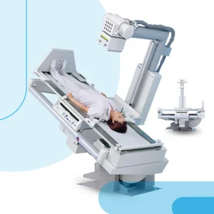
Fluoroscopy is a medical imaging technique that utilizes X-rays to generate real-time moving images of the internal structures of a patient’s body. It operates based on the principle of X-ray transmission through the body, where a continuous beam of X-rays is directed towards the patient. These X-rays pass through the body and are attenuated to varying degrees by different tissues and structures based on their density and composition. The attenuated X-rays are then captured by a detector, which converts them into electrical signals. These signals are further processed and displayed as a dynamic video image on a monitor in real-time, allowing healthcare professionals to visualize the movement and functionality of organs, tissues, and medical devices such as catheters or contrast agents during various diagnostic or interventional procedures.
The basic principle of fluoroscopy involves the use of a fluoroscope, which typically consists of an X-ray source, a patient table or platform, an image intensifier or flat-panel detector, and a display monitor. The X-ray source emits a controlled beam of X-rays towards the patient, while the image intensifier or detector captures the attenuated X-rays that pass through the patient’s body. The captured X-ray signals are amplified or converted into digital data, which is then processed and displayed as a dynamic video image on the monitor. This real-time imaging capability allows healthcare professionals to perform procedures such as gastrointestinal studies, angiography, cardiac catheterization, and orthopedic interventions with precision and accuracy.
Fluoroscopy finds extensive applications in various medical specialties, including surgery, interventional radiology, cardiology, orthopedics, and gastroenterology. It plays a crucial role in guiding and monitoring procedures such as endoscopic retrograde cholangiopancreatography (ERCP), barium studies, cardiac catheterization, joint injections, and spine interventions. The dynamic nature of fluoroscopic imaging enables healthcare providers to visualize anatomical structures in motion, assess physiological functions, and guide therapeutic interventions in real-time, enhancing diagnostic accuracy and treatment outcomes.
The advantages of fluoroscopy include its ability to provide real-time, dynamic images that enable immediate assessment and intervention during procedures. It offers high-resolution imaging with enhanced contrast and detail, allowing for precise visualization of anatomical structures and pathological conditions. Fluoroscopy also facilitates minimally invasive procedures by guiding catheters, needles, and other medical instruments to target areas with accuracy and efficiency, reducing the risk of complications and improving patient outcomes.
Safety measures are paramount in fluoroscopy to minimize radiation exposure to patients and healthcare providers. Techniques such as pulsed fluoroscopy, dose modulation, collimation, and beam filtration are employed to optimize image quality while minimizing radiation dose. Lead aprons, thyroid shields, and other protective equipment are used to shield healthcare personnel from radiation exposure during fluoroscopic procedures. Additionally, strict adherence to radiation safety protocols, dose monitoring, and regular equipment maintenance are essential to ensure safe and effective use of fluoroscopy in clinical practice.
Patient rights and preferences are central to the fluoroscopy process, and informed consent is obtained before performing any fluoroscopic procedure. Patients have the right to be informed about the risks, benefits, and alternatives of the procedure, as well as the expected outcomes and potential complications. They should also be given the opportunity to ask questions, express concerns, and participate in decision-making regarding their healthcare. Respecting patient privacy, dignity, and autonomy is crucial throughout the fluoroscopy examination, and efforts should be made to ensure their comfort and well-being during the procedure.
Technological advancements continue to drive innovation in fluoroscopy, with ongoing developments aimed at improving image quality, reducing radiation dose, and enhancing procedural efficiency. Digital fluoroscopy systems with flat-panel detectors offer higher spatial resolution, faster image acquisition, and lower radiation dose compared to traditional image intensifier-based systems. Advanced image processing techniques, such as digital subtraction angiography (DSA) and three-dimensional (3D) reconstruction, further enhance the diagnostic capabilities of fluoroscopy by providing detailed anatomical information and improving lesion detection.
Looking ahead, the future of fluoroscopy is likely to be shaped by advancements in digital imaging technology, artificial intelligence (AI), and image-guided interventions. Integration of AI algorithms for image analysis and procedural guidance holds promise for improving diagnostic accuracy, reducing procedure times, and optimizing patient outcomes in fluoroscopic procedures. Additionally, emerging technologies such as augmented reality (AR) and virtual reality (VR) may offer new opportunities for immersive visualization and training in fluoroscopy, further advancing its clinical utility and educational value.
In summary, fluoroscopy is a versatile and indispensable imaging modality that enables real-time visualization of anatomical structures and dynamic physiological processes. With ongoing technological advancements and adherence to patient-centered care principles, fluoroscopy continues to play a vital role in modern healthcare, facilitating accurate diagnosis, guiding therapeutic interventions, and improving patient outcomes across a wide range of medical specialties.
Basic Principles and Principles of Fluoroscopy
The basic principle of fluoroscopy involves the use of X-rays to create real-time moving images of internal structures within the body. This imaging technique relies on the differential attenuation of X-rays by various tissues and structures based on their density and composition. When an X-ray beam passes through the body, it is attenuated to varying degrees by different tissues, with denser structures absorbing more X-rays and appearing brighter on the fluoroscopic image. The attenuated X-rays are then captured by a detector, which converts them into electrical signals. These signals are further processed and displayed as a dynamic video image on a monitor in real-time, allowing healthcare professionals to visualize the movement and functionality of organs, tissues, and medical devices during diagnostic or interventional procedures.
During fluoroscopy scanning, patients may experience various sensations depending on the specific procedure being performed and their individual medical condition. Some common aspects of the patient experience during fluoroscopy include:
Preparation: Patients may be required to remove clothing or jewelry that could interfere with the imaging process. They may also need to fast for a certain period before the procedure, especially if contrast agents will be used.
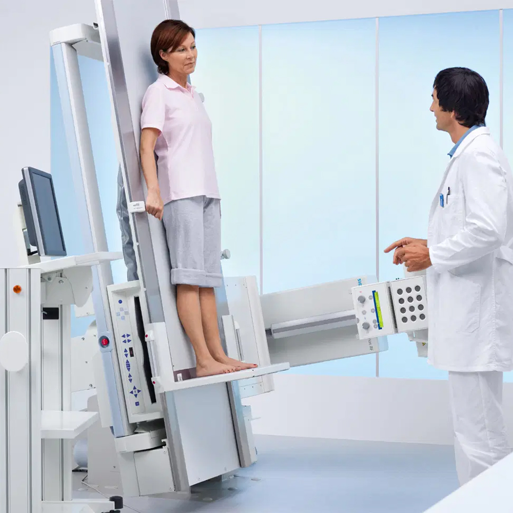
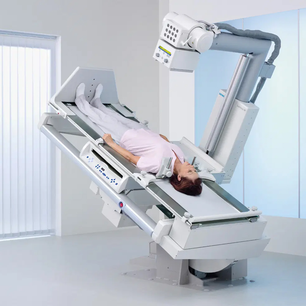
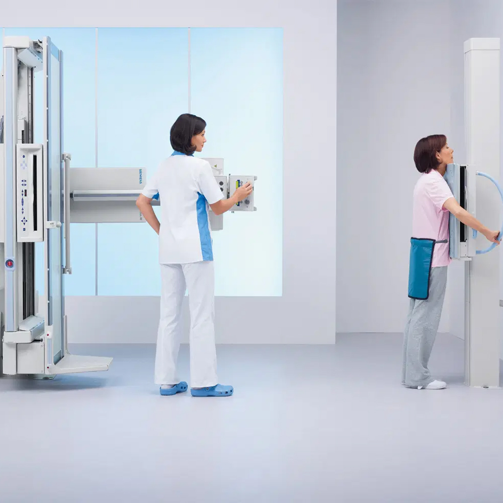
Positioning: Patients are positioned on an examination table or fluoroscopy table in a way that allows the area of interest to be adequately visualized. This positioning may involve lying down, sitting, or standing, depending on the type of procedure.
Comfort: It is essential for patients to remain as comfortable as possible during the procedure, which may involve staying still for extended periods. Pillows or supportive devices may be used to help patients maintain the required position and minimize discomfort.
Communication: Healthcare professionals performing the fluoroscopy procedure will communicate with patients throughout the process to ensure their comfort and cooperation. Patients may be instructed to hold their breath or make specific movements as needed to obtain optimal imaging.
Duration: The duration of fluoroscopy scanning can vary depending on the complexity of the procedure and the area being examined. Some procedures may be relatively brief, while others may require more extended periods of imaging.
Radiation exposure: Patients may have concerns about radiation exposure during fluoroscopy scanning. Healthcare providers take measures to minimize radiation exposure by using the lowest possible dose of radiation necessary to obtain the required images.
Monitoring: Patients are continuously monitored during fluoroscopy scanning to ensure their safety and well-being. Healthcare professionals closely observe vital signs and any signs of discomfort or adverse reactions throughout the procedure.
Overall, the patient experience during fluoroscopy scanning aims to prioritize comfort, safety, and effective communication between the patient and healthcare providers to ensure the successful completion of the procedure.
Fluoroscopy is a versatile imaging technique used in various medical specialties for both diagnostic and interventional purposes. Its applications and uses span across several medical fields, including:
Diagnostic Imaging: Fluoroscopy is commonly used to visualize the movement and functionality of internal organs and structures in real-time. It is particularly valuable in procedures such as gastrointestinal studies (barium swallow, barium enema), urological studies (voiding cystourethrography), and musculoskeletal evaluations (joint arthrography).
Interventional Procedures: Fluoroscopy-guided interventions are performed for a wide range of purposes, including minimally invasive surgeries, pain management procedures (e.g., epidural steroid injections), orthopedic procedures (e.g., joint injections), and vascular interventions (e.g., angiography, catheter-based treatments).
Orthopedics: Fluoroscopy is extensively used in orthopedic procedures for guiding fracture reductions, joint injections, arthrograms, and spinal interventions such as vertebroplasty and kyphoplasty.
Cardiology: In cardiology, fluoroscopy is utilized for procedures such as cardiac catheterization, electrophysiology studies, pacemaker implantation, and coronary angiography.
Pulmonology: Fluoroscopy plays a role in pulmonary procedures, including bronchoscopy and placement of chest tubes.
Vascular Surgery: Fluoroscopy is integral to vascular interventions, including angioplasty, stent placement, embolization, and thrombolysis.
Gastroenterology: In gastroenterology, fluoroscopy aids in procedures such as endoscopic retrograde cholangiopancreatography (ERCP), balloon enteroscopy, and percutaneous gastrostomy tube placement.
Image Acquisition and Processing: Fluoroscopy involves the real-time acquisition of X-ray images, which are continuously displayed on a monitor. The images are captured using an X-ray source and a fluoroscopic detector, typically a flat-panel detector or an image intensifier. The acquired images are processed in real-time to enhance image quality, adjust contrast and brightness, and reduce noise, providing clear visualization of anatomical structures and dynamic movements.
Patient Preparation and Experience During Fluoroscopy Scanning: Patient preparation for fluoroscopy varies depending on the specific procedure being performed. It may involve fasting before the procedure, removing clothing or jewelry that could interfere with imaging, and discussing any relevant medical history or allergies with the healthcare team. During the fluoroscopy procedure, patients are positioned on an examination table or fluoroscopy table as needed, and they may be asked to hold their breath or change positions to facilitate imaging. Communication with the healthcare team is essential to ensure patient comfort and cooperation throughout the procedure. Patients may experience minimal discomfort or anxiety related to the procedure, but healthcare providers strive to minimize this through effective communication, supportive positioning, and ensuring a safe and efficient imaging process.
Pre-Scanning Preparation Process: Before undergoing a fluoroscopy procedure, patients may be instructed to follow specific preparation guidelines depending on the type of examination or intervention planned. This may include fasting for a certain period if the procedure involves the digestive system, removing metallic objects or jewelry that may interfere with imaging, and informing the healthcare team about any allergies or medical conditions. Additionally, patients may receive instructions regarding medication use, such as whether to continue taking prescribed medications or to temporarily discontinue certain medications prior to the procedure.
Patient Comfort and Convenience During Scanning: Ensuring patient comfort and convenience during fluoroscopy scanning is crucial for a successful procedure. Healthcare providers strive to create a comfortable environment for patients, addressing any concerns they may have and providing support throughout the process. Patients are positioned comfortably on the examination table or fluoroscopy table, and measures are taken to minimize discomfort during the procedure. Clear communication between the healthcare team and the patient helps to alleviate any anxiety or apprehension, and patients are encouraged to ask questions or express any discomfort they may experience during the procedure.
Advantages and Benefits of Fluoroscopy: Fluoroscopy offers several advantages and benefits over traditional radiographic imaging, including:
Real-time Imaging: Fluoroscopy provides dynamic, real-time imaging of anatomical structures and physiological processes, allowing healthcare providers to visualize movements and changes as they occur.
Enhanced Visualization: Fluoroscopy enables enhanced visualization of soft tissues, organs, and structures, making it particularly valuable for guiding interventional procedures and assessing functional abnormalities.
Minimally Invasive Procedures: Fluoroscopy-guided interventions allow for minimally invasive procedures, reducing the need for traditional open surgeries and resulting in shorter recovery times, less postoperative pain, and reduced risk of complications.
Immediate Feedback: The real-time nature of fluoroscopy provides immediate feedback to healthcare providers during procedures, allowing for prompt adjustments and interventions as needed to ensure optimal outcomes.
Versatility: Fluoroscopy is versatile and can be used across various medical specialties, including cardiology, gastroenterology, orthopedics, pulmonology, urology, and vascular surgery, among others.
Real-Time Imaging and Dynamic Capabilities: One of the primary advantages of fluoroscopy is its real-time imaging capabilities, which allow healthcare providers to observe dynamic processes and movements within the body as they occur. This real-time feedback is invaluable for guiding interventional procedures, such as catheter-based treatments, joint injections, and gastrointestinal studies, where precise positioning and visualization are essential for successful outcomes. Additionally, fluoroscopy’s dynamic capabilities enable healthcare providers to assess functional abnormalities, such as swallowing disorders or urinary tract obstructions, by visualizing the movement and function of affected structures in real-time.
Ease of Use in Diagnostic and Interventional Procedures: Fluoroscopy is highly valued for its ease of use in both diagnostic imaging and interventional procedures. Its real-time imaging capabilities provide immediate feedback to healthcare providers during procedures, allowing for precise guidance and accurate placement of instruments or devices. This ease of use enhances the efficiency and accuracy of diagnostic examinations and minimally invasive interventions, leading to improved patient outcomes and reduced procedural times.
Safety of Fluoroscopy Scanning: Ensuring the safety of patients during fluoroscopy scanning is paramount. Healthcare providers adhere to strict safety protocols to minimize radiation exposure and mitigate associated risks. This includes employing low-dose fluoroscopy techniques whenever possible, limiting the duration of exposure, and utilizing protective shielding to minimize radiation exposure to non-targeted areas of the body. Additionally, continuous monitoring of radiation dose levels and adherence to ALARA (As Low As Reasonably Achievable) principles are integral to ensuring patient safety during fluoroscopy procedures.
Evaluation and Interpretation of Fluoroscopy Images: The evaluation and interpretation of fluoroscopy images require specialized training and expertise. Healthcare providers carefully analyze fluoroscopic images to assess anatomical structures, identify abnormalities or pathology, and guide diagnostic and interventional decision-making. Interpretation may involve assessing the size, shape, and position of structures, as well as evaluating the dynamics of physiological processes captured in real-time. Clear communication between radiologists, interventionalists, and other healthcare team members is essential for accurate image interpretation and optimal patient care.
Understanding Image Results and Sharing with the Doctor: After fluoroscopy imaging is performed, healthcare providers review and interpret the results to formulate diagnostic impressions and treatment plans. Patients may be involved in discussions about their imaging findings, and healthcare providers ensure that the results are explained in understandable terms. Clear communication between the healthcare team and the patient facilitates patient understanding of the imaging results and the implications for their health. Additionally, imaging results are shared with referring physicians or specialists to facilitate collaborative decision-making and ongoing patient care.
Technological Developments and Innovations in Fluoroscopy: Advancements in fluoroscopy technology have led to the development of various innovative features and capabilities. These include improvements in image quality through higher resolution detectors and advanced image processing algorithms, enhancing visualization of anatomical structures and pathology. Additionally, the integration of advanced software solutions enables real-time image enhancement and manipulation, facilitating more accurate diagnosis and treatment planning. Furthermore, advancements in radiation dose reduction techniques, such as pulsed fluoroscopy and dose modulation algorithms, contribute to improved patient safety during fluoroscopic procedures.
Improvements in Fluoroscopy Techniques and Future Potential: Continuous research and development efforts aim to enhance fluoroscopy techniques and expand their clinical utility. Future advancements may include the integration of artificial intelligence (AI) algorithms for real-time image analysis and decision support, improving diagnostic accuracy and efficiency. Furthermore, the development of novel contrast agents and imaging protocols may enable enhanced visualization of specific anatomical structures and physiological processes, further advancing the diagnostic capabilities of fluoroscopy. Additionally, advancements in miniaturized fluoroscopy systems and portable devices may facilitate point-of-care imaging in various clinical settings, increasing accessibility and expanding the reach of fluoroscopy technology.
Patient Rights and Information About Fluoroscopy Scanning: Patients undergoing fluoroscopy procedures have the right to be informed about the nature of the procedure, its purpose, potential risks, and alternative options. Healthcare providers must obtain informed consent from patients before performing fluoroscopy examinations, ensuring that patients are aware of the benefits and risks associated with the procedure. Additionally, patients have the right to privacy and confidentiality regarding their medical information, and healthcare providers must adhere to strict data protection regulations to safeguard patient data collected during fluoroscopy procedures.
Privacy and Data Protection: Privacy and data protection are critical considerations in fluoroscopy imaging to ensure patient confidentiality and compliance with regulatory requirements. Healthcare providers must implement robust data security measures to protect patient information collected during fluoroscopy procedures, including encryption, access controls, and secure storage protocols. Additionally, adherence to relevant privacy laws and regulations, such as the Health Insurance Portability and Accountability Act (HIPAA) in the United States, is essential to safeguard patient privacy rights and prevent unauthorized access or disclosure of sensitive medical information.
Patient Rights and Preferences During the Scanning Process: During fluoroscopy procedures, patients have specific rights and preferences that must be respected by healthcare providers. These include the right to be informed about the procedure’s purpose, risks, and potential benefits, as well as any alternative options available. Patients also have the right to actively participate in decision-making regarding their healthcare, including the choice to consent or refuse the procedure. Furthermore, healthcare providers should prioritize patient comfort and address any concerns or preferences related to the scanning process, such as the use of sedation or anesthesia, positioning during the procedure, and communication with the medical team.
Future and Development of Fluoroscopy Applications: The future of fluoroscopy applications is characterized by ongoing advancements aimed at expanding its clinical utility and improving patient outcomes. Emerging technologies and innovative techniques are being developed to enhance the diagnostic and interventional capabilities of fluoroscopy across various medical specialties. Future developments may include the integration of artificial intelligence (AI) and machine learning algorithms to automate image analysis, improve procedural guidance, and personalize treatment approaches. Furthermore, advancements in imaging hardware and software are expected to enable higher resolution imaging, faster image acquisition, and enhanced visualization of anatomical structures and pathology. Additionally, the integration of fluoroscopy with other imaging modalities, such as computed tomography (CT) and magnetic resonance imaging (MRI), may further expand its applications and clinical impact.
Advancements in Fluoroscopy and Future Directions: Advancements in fluoroscopy technology are driving significant improvements in diagnostic and interventional procedures, with ongoing developments shaping its future directions. Key areas of advancement include radiation dose reduction techniques to enhance patient safety, such as pulsed fluoroscopy and dose modulation algorithms. Additionally, improvements in image quality and resolution are facilitating more accurate diagnosis and treatment planning. Future directions may also include the development of miniaturized fluoroscopy systems for portable and point-of-care imaging, as well as the integration of advanced imaging guidance techniques for complex interventions. Furthermore, advancements in fluoroscopic imaging software and hardware are expected to enable real-time image fusion, augmented reality visualization, and enhanced procedural guidance, further optimizing clinical outcomes and patient care.
Frequently Asked Questions
Get a Free Second Opinion
Experienced Burtom Medical Team is Ready to Help
I consent to Burtom Health Group using my aforesaid personal data for the purposes described in this notice and understand that I can withdraw my consent at any time by sending a request to info@burtom.com.


