Burtom ‣ Technologies ‣ Mobile C-arm System (Fluoroscopy)
Language: 🇬🇧 English | 🇹🇷 Türkçe
Mobile C-arm System (Fluoroscopy) Overview

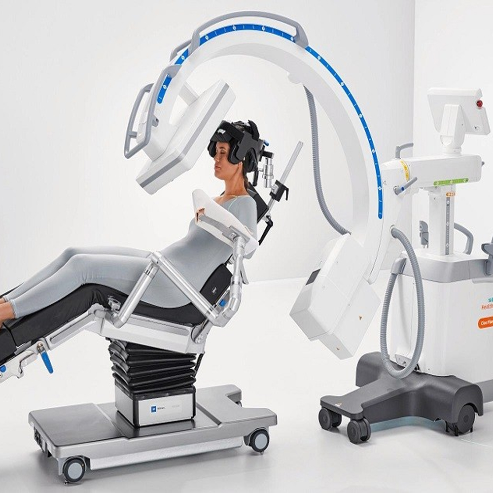
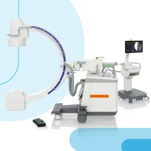 The mobile C-arm system is a sophisticated medical imaging device that combines X-ray fluoroscopy and radiography capabilities in a single unit. It consists of a C-shaped arm with an X-ray source on one end and an image detector on the other, connected by a mobile gantry. This design allows for flexible positioning and movement around the patient during surgical and interventional procedures.
One of the primary uses of the mobile C-arm system is in orthopedic surgery, where it provides real-time imaging of bones and joints to assist in procedures such as fracture reduction, fixation, and joint injections. It is also commonly used in vascular, cardiac, and neurosurgical procedures for visualizing catheter placement, blood flow, and soft tissue anatomy.
The system operates by emitting X-ray radiation from the source, which passes through the patient and is detected by the image receptor. The resulting images are displayed in real-time on a monitor, allowing the surgeon or interventionalist to visualize the internal structures of the patient’s body as they perform the procedure. This real-time imaging capability enables precise navigation and immediate feedback, leading to more accurate and effective treatments.
In addition to its utility in the operating room, the mobile C-arm system is also used in other medical settings such as emergency departments, intensive care units, and outpatient clinics. Its portability and ease of use make it a valuable tool for imaging patients at the point of care, without the need for transporting them to a dedicated radiology suite.
Overall, the mobile C-arm system plays a crucial role in modern healthcare by providing high-quality imaging during surgical and interventional procedures, enhancing patient care, and improving clinical outcomes. Its versatility, mobility, and real-time imaging capabilities make it an indispensable tool for medical professionals across various specialties.

Mobile C-Arm System (Fluoroscopy) Description and Function
Mobile C-arm systems are medical imaging devices primarily used for fluoroscopy-guided procedures in various medical specialties such as orthopedics, vascular surgery, and interventional radiology. These systems consist of a C-shaped arm with an X-ray source on one end and an image intensifier or flat-panel detector on the other end. The C-arm can be easily maneuvered around the patient, providing real-time X-ray images of the targeted area during surgical or interventional procedures. This enables clinicians to visualize internal structures, guide surgical instruments, and monitor treatment progress with precision and efficiency. Mobile C-arm systems play a crucial role in improving procedural outcomes, reducing surgical time, and minimizing patient radiation exposure compared to traditional surgical methods.
Basic Principles and Applications of the Mobile C-Arm System
Mobile C-arm systems are pivotal tools in various medical settings, offering real-time imaging capabilities for a wide array of procedures. These systems operate based on X-ray technology, utilizing a C-shaped arm that can be easily maneuvered around the patient. The basic principle involves emitting X-rays from one side of the C-arm, which then pass through the patient’s body and are captured by an image receptor on the opposite side. This process creates real-time images that assist healthcare professionals during surgical, orthopedic, and interventional procedures.
Importance of Mobility and Portability
The mobility and portability of C-arm systems significantly contribute to their versatility and effectiveness in medical practice. Unlike traditional fixed imaging systems, mobile C-arms can be easily transported within healthcare facilities to various departments or even between different medical facilities. This flexibility allows for on-the-spot imaging during surgeries, emergency interventions, or bedside procedures, reducing the need for patient transportation and minimizing procedural delays. Additionally, the compact design of mobile C-arm systems ensures accessibility in confined spaces such as operating rooms, emergency departments, or outpatient clinics, further enhancing their utility in diverse medical settings.
Applications of the Mobile C-Arm System
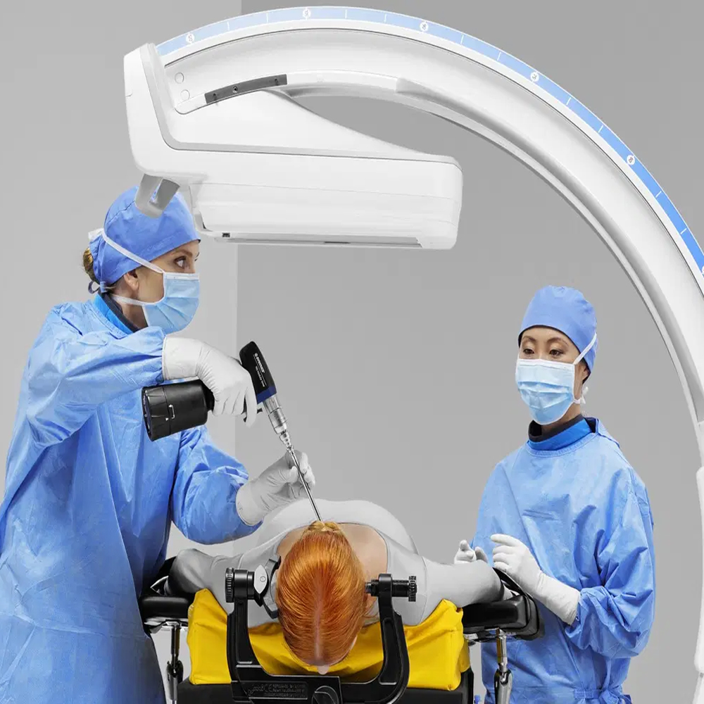
Mobile C-arm systems find extensive applications across a spectrum of medical specialties, including orthopedics, trauma surgery, vascular procedures, and pain management interventions. In orthopedic surgeries, these systems facilitate real-time visualization of bone structures, fracture reduction, and implant placement, aiding surgeons in achieving optimal surgical outcomes. Furthermore, mobile C-arms play a crucial role in trauma surgery by providing rapid imaging capabilities for assessing traumatic injuries and guiding surgical interventions in emergency settings. Additionally, in vascular procedures, these systems enable precise localization of catheters and guidewires during angiography or endovascular interventions, ensuring accurate placement and enhancing procedural success rates. Moreover, mobile C-arms are valuable tools in pain management interventions such as fluoroscopic-guided injections or nerve blocks, allowing for precise needle placement and targeted delivery of therapeutic agents to alleviate pain.
Technological Features of the Mobile C-Arm System
The mobile C-arm system incorporates advanced technological features that enhance its imaging capabilities and operational efficiency. These systems are equipped with high-frequency X-ray generators and digital flat-panel detectors, enabling superior image quality with reduced radiation dose. Moreover, modern mobile C-arms often feature pulse fluoroscopy and pulsed fluoroscopy modes, allowing for dynamic imaging while minimizing radiation exposure to patients and healthcare personnel. Additionally, many systems are equipped with advanced image processing software, offering features such as noise reduction, edge enhancement, and real-time image stitching for seamless visualization of large anatomical regions.
Basic Components and Operation of the Mobile C-Arm System
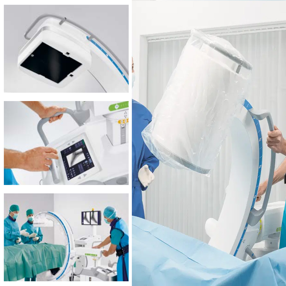
The mobile C-arm system consists of several essential components that facilitate its operation and image acquisition. The primary components include the C-shaped gantry, X-ray tube, image intensifier or flat-panel detector, control panel, and monitor display. The C-shaped gantry houses the X-ray tube and detector assembly, which can be maneuvered into various positions around the patient during imaging procedures. The X-ray tube emits X-rays, which pass through the patient’s body and are captured by the image intensifier or flat-panel detector. The control panel allows the technologist or surgeon to adjust imaging parameters such as exposure settings, pulse rate, and image processing options. The monitor display provides real-time visualization of acquired images, enabling immediate assessment and guidance during procedures.
Image Quality and Resolution
Image quality and resolution are critical factors in mobile C-arm imaging systems, as they directly impact the accuracy and diagnostic value of acquired images. Modern mobile C-arms utilize digital flat-panel detectors, which offer superior image quality compared to traditional image intensifiers. These detectors provide high spatial resolution and dynamic range, allowing for detailed visualization of anatomical structures with excellent contrast and clarity. Furthermore, advanced image processing algorithms enhance image quality by reducing noise, optimizing contrast, and improving edge definition. The high-resolution images produced by mobile C-arm systems enable precise anatomical localization, accurate assessment of pathology, and effective guidance during interventional procedures, ultimately contributing to improved patient outcomes.
Safe Use of the Mobile C-Arm System
Ensuring the safe use of the mobile C-arm system is paramount to protect both patients and healthcare personnel from potential hazards associated with radiation exposure. Healthcare facilities must adhere to strict safety protocols and guidelines to minimize risks during imaging procedures. Technologists and physicians operating the mobile C-arm should receive comprehensive training on radiation safety practices, including proper positioning of the equipment, optimization of imaging parameters, and effective use of protective shielding devices.
Radiation Exposure and Safety Measures
Mobile C-arm systems utilize ionizing radiation to generate medical images, posing potential risks of radiation exposure to patients and healthcare providers. To mitigate these risks, stringent safety measures are implemented to limit radiation exposure levels while maintaining diagnostic image quality. These measures include adhering to the “as low as reasonably achievable” (ALARA) principle, which emphasizes minimizing radiation dose without compromising diagnostic efficacy. Additionally, personnel should use lead aprons, thyroid shields, and protective eyewear to reduce radiation exposure during imaging procedures. Furthermore, continuous monitoring of radiation dose levels and regular equipment maintenance are essential to ensure compliance with safety standards and regulatory requirements.
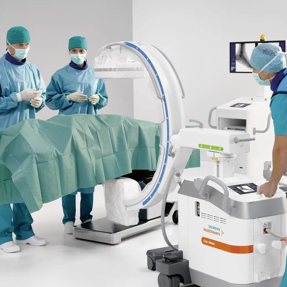
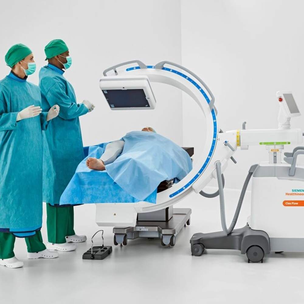
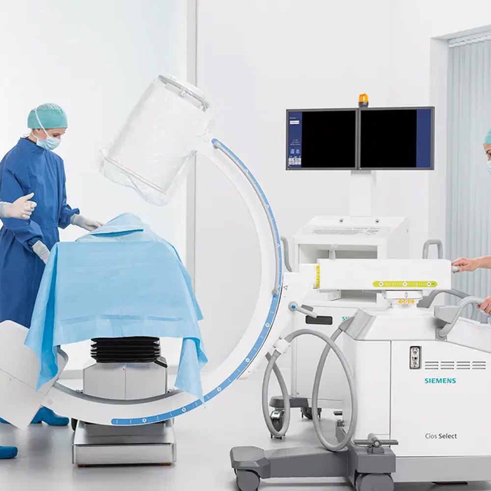
Patient and Staff Safety During Surgical Procedures
During surgical procedures performed under mobile C-arm guidance, ensuring patient and staff safety is of utmost importance. Healthcare providers must take precautions to minimize radiation exposure while maintaining optimal imaging quality. This involves proper positioning of the C-arm to achieve optimal imaging angles while maximizing distance from the radiation source. Additionally, personnel should wear appropriate protective equipment, including lead aprons, thyroid shields, and leaded glasses, to reduce radiation exposure. Furthermore, effective communication between surgical team members is crucial to coordinate movements and minimize unnecessary exposure to radiation. By implementing comprehensive safety protocols and adhering to radiation safety guidelines, healthcare facilities can ensure the safe use of mobile C-arm systems during surgical procedures, safeguarding the well-being of both patients and staff.
Benefits and Advantages of the Mobile C-Arm System
The mobile C-arm system offers numerous benefits and advantages in various medical settings, particularly during surgical interventions. One of the key advantages is its portability, allowing for flexibility in positioning and maneuverability within different clinical environments. This mobility enables real-time imaging during surgical procedures, facilitating accurate intraoperative guidance and decision-making.
Precise and Rapid Results in Surgical Interventions
The mobile C-arm system provides surgeons with immediate access to high-quality imaging, allowing for precise visualization of anatomical structures during surgical interventions. With real-time imaging capabilities, surgeons can assess the progress of procedures, verify instrument placement, and make necessary adjustments instantly. This rapid feedback loop enhances procedural efficiency, reduces operative time, and minimizes the risk of intraoperative complications.
Applications and Case Examples of the Mobile C-Arm System
The mobile C-arm system finds extensive applications across various medical specialties, including orthopedics, trauma surgery, interventional radiology, and cardiovascular procedures. In orthopedic surgery, it is commonly used for intraoperative imaging of fractures, joint replacements, and spinal procedures. Trauma surgeons rely on mobile C-arm imaging to assess and manage acute injuries in emergency settings.
In interventional radiology, the mobile C-arm system plays a crucial role in minimally invasive procedures such as angiography, embolization, and percutaneous interventions. Additionally, it is utilized in cardiovascular procedures such as cardiac catheterization and electrophysiology studies to guide catheter placement and visualize cardiac anatomy.
Case examples of the mobile C-arm system’s applications include intraoperative localization of foreign bodies, placement of orthopedic implants, guidance for spinal fusion surgeries, and visualization of vascular interventions. By providing real-time imaging guidance, the mobile C-arm system enhances procedural accuracy, improves patient outcomes, and contributes to the overall efficiency of surgical interventions.
Use in Cardiovascular and Orthopedic Surgery
The mobile C-arm system plays a pivotal role in cardiovascular and orthopedic surgery, offering real-time imaging capabilities that aid in intricate procedures. In cardiovascular surgery, it facilitates the visualization of blood vessels and cardiac structures during interventions such as angiography, stent placement, and pacemaker implantation. Orthopedic surgeons utilize the system for intraoperative imaging of fractures, joint replacements, and spinal surgeries, ensuring precise implant placement and alignment.
Limitations of Mobile C-Arm System Use
Despite its versatility, the mobile C-arm system has certain limitations that need to be considered. One limitation is its inability to provide three-dimensional (3D) imaging, which may be necessary for complex anatomical assessments or preoperative planning. Additionally, the system’s image quality may be affected by patient size, tissue density, and the presence of artifacts, leading to suboptimal visualization in some cases. Furthermore, the mobility of the system may be restricted in crowded operating rooms or tight spaces, limiting its positioning and imaging capabilities.
Challenges in Imaging Deep Organs
Imaging deep organs poses a challenge for the mobile C-arm system due to its limited penetration depth and potential for image distortion. Deeply situated anatomical structures may not be adequately visualized, especially in obese patients or those with thick tissue layers. Additionally, artifacts caused by overlying bones or metal implants can obscure the visibility of deep organs, necessitating alternative imaging modalities or intraoperative techniques for optimal visualization. Despite these challenges, advancements in imaging technology and surgical techniques continue to improve the system’s efficacy in imaging deep organs during surgical procedures.
Comparison with Other Imaging Techniques for Some Diseases
When compared to other imaging techniques, the mobile C-arm system offers distinct advantages in certain clinical scenarios. For instance, in trauma cases involving skeletal injuries, such as fractures or dislocations, the real-time imaging capabilities of the C-arm allow for immediate assessment and guidance during reduction procedures, leading to improved patient outcomes. Similarly, in interventional procedures such as percutaneous transluminal angioplasty (PTA) or embolization, the dynamic imaging provided by the C-arm enables precise catheter navigation and intervention delivery, reducing procedure times and minimizing patient discomfort compared to conventional angiography suites.
Patient Rights and Important Considerations
Ensuring patient rights and considerations are paramount when utilizing the mobile C-arm system. Patients have the right to be informed about the procedure, including its risks, benefits, and alternatives, allowing them to make informed decisions. Additionally, patient privacy and dignity must be upheld throughout the imaging process, with appropriate measures taken to minimize exposure and maintain confidentiality. Informed consent should be obtained before the procedure, detailing the purpose, potential risks, and expected outcomes. Moreover, patient comfort and safety should be prioritized, with adequate pain management and monitoring during the imaging procedure.
Privacy and Data Protection
Maintaining privacy and data protection is essential when using the mobile C-arm system, as it involves the acquisition and storage of patient imaging data. Healthcare facilities must adhere to strict guidelines and regulations, such as the Health Insurance Portability and Accountability Act (HIPAA) in the United States, to safeguard patient information. This includes implementing secure data storage and transmission protocols, restricting access to authorized personnel only, and ensuring encryption of sensitive data. Regular audits and assessments of data security measures should be conducted to identify and address any vulnerabilities, thereby maintaining patient privacy and data protection.
Technological Developments and Future Directions
The field of mobile C-arm systems is witnessing rapid technological advancements, paving the way for innovative applications and improved patient care. One key area of development is the integration of advanced imaging modalities, such as 3D reconstruction and augmented reality, into mobile C-arm systems. These enhancements enable more precise anatomical visualization and enhanced surgical guidance, leading to better treatment outcomes.
Advancements in Hardware and Software
Hardware and software advancements play a crucial role in enhancing the capabilities of mobile C-arm systems. On the hardware front, developments in X-ray tube technology, detector design, and mechanical components have led to improvements in image quality, dose efficiency, and system maneuverability. Meanwhile, software enhancements, including real-time image processing algorithms and user-friendly interfaces, facilitate efficient image acquisition, manipulation, and interpretation, thereby streamlining surgical workflows.
Potential of Mobile C-Arm Systems in the Future
The future of mobile C-arm systems holds great promise across various medical specialties and clinical settings. These systems are expected to become more compact, lightweight, and portable, enabling their deployment in a wider range of healthcare facilities, including ambulatory surgical centers and remote clinics. Additionally, advancements in miniaturization and wireless connectivity may facilitate the development of wearable or handheld C-arm devices, further expanding their utility in point-of-care settings and intraoperative imaging.
Moreover, the integration of artificial intelligence (AI) and machine learning algorithms into mobile C-arm systems holds immense potential for enhancing diagnostic accuracy, surgical planning, and intraoperative decision-making. AI-powered image analysis tools can assist clinicians in identifying abnormalities, measuring anatomical parameters, and predicting treatment outcomes, ultimately improving patient care and surgical precision.
Overall, the ongoing technological developments and future directions in mobile C-arm systems are poised to revolutionize intraoperative imaging and surgical interventions, offering clinicians advanced capabilities and patients better treatment options.
Future Applications and Development of the Mobile C-Arm System
The future of mobile C-arm systems is characterized by an array of exciting developments and potential applications that are set to transform intraoperative imaging and surgical interventions. One significant area of advancement is the integration of emerging technologies, such as artificial intelligence (AI) and augmented reality (AR), into mobile C-arm systems. These technologies have the potential to enhance surgical precision, improve patient outcomes, and streamline clinical workflows.
New Technologies and Application Areas
Emerging technologies, including AI-based image processing algorithms and deep learning models, are expected to play a crucial role in the evolution of mobile C-arm systems. These technologies can facilitate real-time image analysis, automated segmentation of anatomical structures, and decision support for surgeons during complex procedures. Additionally, advancements in imaging modalities, such as cone-beam CT (CBCT) and dual-energy imaging, may expand the diagnostic capabilities of mobile C-arm systems, enabling more comprehensive intraoperative assessment of soft tissue and bone structures.
Furthermore, the integration of AR-based surgical navigation systems with mobile C-arm units holds promise for enhancing surgical precision and accuracy. AR overlays can provide surgeons with real-time anatomical guidance, intraoperative visualization of critical structures, and procedural feedback, leading to improved surgical outcomes and reduced complications.
Development and Improvement Potential of Mobile C-Arm Systems
The development and improvement potential of mobile C-arm systems extend beyond technological advancements to include ergonomic design enhancements, user-friendly interfaces, and seamless integration with surgical workflows. Future iterations of mobile C-arm systems may feature compact, lightweight designs with enhanced maneuverability and ergonomic controls, allowing for easier positioning and manipulation during surgical procedures.
Moreover, efforts to optimize dose efficiency and image quality while minimizing radiation exposure to patients and healthcare personnel will continue to drive advancements in mobile C-arm technology. Innovations in X-ray tube design, detector technology, and dose management algorithms aim to achieve higher spatial resolution, improved contrast resolution, and lower radiation doses, ensuring the safety and well-being of both patients and surgical staff.
In summary, the future applications and development of mobile C-arm systems are characterized by the integration of advanced technologies, expansion of imaging modalities, and refinement of system design and usability. These advancements hold the potential to revolutionize intraoperative imaging and surgical interventions, enabling safer, more efficient, and more precise patient care across a wide range of medical specialties.
Frequently Asked Questions
Get a Free Second Opinion
Experienced Burtom Medical Team is Ready to Help
I consent to Burtom Health Group using my aforesaid personal data for the purposes described in this notice and understand that I can withdraw my consent at any time by sending a request to info@burtom.com.


