Burtom ‣ Technologies ‣ Open Magnetic Resonance 0.35 Tesla (MRI)
Language: 🇬🇧 English | 🇹🇷 Türkçe
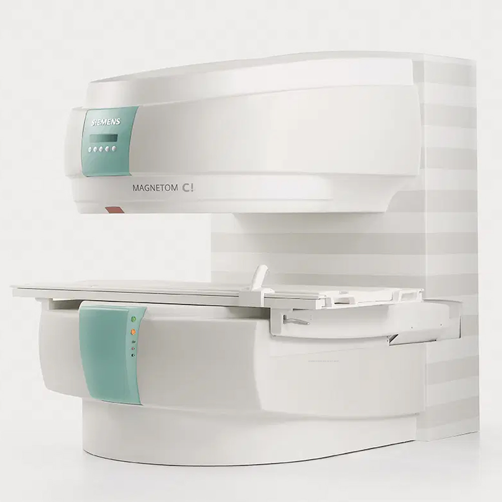
Open Magnetic Resonance 0.35 Tesla (MRI) Overview

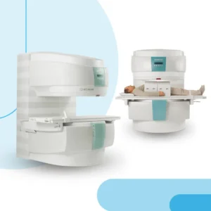 Open Magnetic Resonance 0.35 Tesla (MRI) is a cutting-edge imaging system that utilizes a magnetic field strength of 0.35 Tesla to deliver precise diagnostic imaging. Unlike traditional closed MRI machines, Open MRI 0.35T features an open design, providing patients with a more spacious and less confining scanning experience. This innovative configuration is particularly beneficial for individuals who may feel claustrophobic or uncomfortable in enclosed spaces.
Despite its open design, Open MRI 0.35T does not compromise on imaging quality. It is equipped with advanced technology that ensures high-resolution images, allowing for accurate diagnostic evaluations across various medical applications. From routine scans to specialized imaging needs, this system delivers reliable results while prioritizing patient comfort.
Open Magnetic Resonance 0.35 Tesla (MRI) represents a patient-centric approach to diagnostic imaging, offering a comfortable and accessible solution for individuals seeking a more relaxed MRI experience without compromising on image quality or diagnostic accuracy.

What is Magnetic Resonance Imaging (MRI) and How Does It Work?
Open Magnetic Resonance Imaging (MRI) is an innovative medical imaging technique that allows for the visualization of internal body structures with remarkable detail. Unlike traditional closed MRI systems characterized by their narrow bore and enclosed design, open MRI machines offer a more spacious and less confining environment.
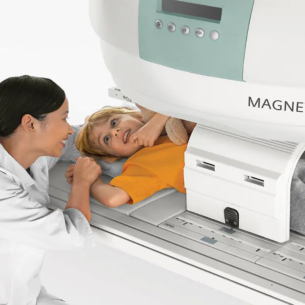
In open MRI setups, patients lie comfortably on a movable table that slides into the open space of the scanner, rather than being confined within a tube. This design is particularly advantageous for individuals who may experience claustrophobia or discomfort in enclosed spaces, as well as those with mobility limitations or larger body sizes.
Despite their open configuration, open MRI systems are capable of producing high-quality images essential for accurate diagnosis and treatment planning across various medical specialties. These systems are utilized to examine a wide range of anatomical regions, including the brain, spine, joints, and abdomen, enabling the detection of diverse medical conditions such as fractures, tumors, and neurological disorders.
In summary, Open Magnetic Resonance Imaging (MRI) offers a patient-centric approach to diagnostic imaging, prioritizing comfort and accessibility without compromising on the quality or accuracy of the imaging results.
The Basic Principle of Open Magnetic Resonance Imaging
The basic principle of Open Magnetic Resonance Imaging (MRI) revolves around the use of magnetic fields and radio waves to generate detailed images of the body’s internal structures. Unlike traditional closed MRI systems, which employ a narrow bore and enclosed design, open MRI machines feature a more spacious and open configuration.
In open MRI systems, the patient lies on a movable table that slides into the open space of the scanner. The scanner consists of powerful magnets that produce a magnetic field around the patient’s body. Radio waves are then directed into the body, causing the hydrogen atoms in the body’s tissues to emit signals. These signals are captured by receivers in the MRI machine and processed by a computer to create detailed cross-sectional images.
The key principle behind open MRI imaging is the interaction between the magnetic field and the body’s hydrogen atoms. By analyzing the signals emitted by these atoms, open MRI systems can generate high-resolution images of the body’s internal structures, providing valuable diagnostic information to healthcare professionals.
Overall, the basic principle of Open Magnetic Resonance Imaging (MRI) involves the use of magnetic fields and radio waves to create detailed images of the body’s anatomy in a comfortable and open environment, facilitating accurate diagnosis and treatment planning.
Advantages of Open Magnetic Resonance Imaging (MRI):
- Enhanced Patient Comfort: The open design of MRI systems reduces feelings of claustrophobia and anxiety, making the scanning experience more comfortable for patients.
- Accommodation for Patients with Mobility Issues: The spacious design of open MRI machines allows for easier access and accommodation of patients with mobility impairments or larger body sizes.
- Pediatric and Geriatric Applications: Open MRI systems are particularly beneficial for pediatric and geriatric patients who may have difficulty remaining still during traditional closed MRI scans.
- Reduced Risk of Sedation: Due to the more accommodating environment, open MRI scans often require less sedation, minimizing associated risks and allowing for safer imaging procedures.
- Improved Patient Communication: The open design enables better communication between healthcare providers and patients during the scanning process, enhancing overall patient experience and compliance.
Applications of Open Magnetic Resonance Imaging (MRI):
- Neurological Imaging: Open MRI systems are utilized for imaging various neurological conditions, including brain tumors, strokes, multiple sclerosis, and traumatic brain injuries.
- Musculoskeletal Imaging: Open MRI is effective for evaluating musculoskeletal disorders such as joint injuries, ligament tears, tendonitis, and osteoarthritis.
- Pediatric Imaging: Open MRI machines are particularly suitable for pediatric imaging due to their accommodating design, enabling the assessment of conditions such as congenital anomalies, developmental disorders, and injuries in children.
- Claustrophobic Patients: Open MRI is an ideal imaging option for patients with claustrophobia or anxiety disorders who may experience distress in traditional closed MRI scanners.
- Geriatric Imaging: Open MRI systems are well-suited for imaging elderly patients, allowing for the assessment of age-related conditions such as degenerative joint diseases, spinal stenosis, and cerebrovascular diseases.
Overall, the advantages and applications of Open Magnetic Resonance Imaging (MRI) make it a valuable tool in diagnostic imaging, providing enhanced patient comfort and accessibility while facilitating accurate diagnosis and treatment across various medical specialties.
Advantages of 0.35 Tesla (0.35T) Magnetic Resonance Imaging (MRI) compared to other magnetic field strengths:
Enhanced Patient Comfort: The lower magnetic field strength of 0.35T MRI systems contributes to a more open and spacious scanning environment, reducing feelings of claustrophobia and discomfort compared to higher field strengths.
Reduced Risk of Artefacts: Lower field strengths, such as 0.35T, are associated with fewer susceptibility artefacts, which can distort images and compromise diagnostic accuracy, particularly in regions near metal implants or air-filled cavities.
Compatibility with Implant Patients: Patients with certain metallic implants or devices may be eligible for imaging with a 0.35T MRI system, as lower field strengths pose less risk of heating or displacement of implants compared to higher field strengths.
Pediatric and Geriatric Applications: 0.35T MRI systems are well-suited for imaging pediatric and geriatric patients due to their reduced noise levels and enhanced patient comfort, making them more tolerable for individuals who may have difficulty remaining still during scans.
Cost-Effectiveness: Lower field strength MRI systems, such as 0.35T, may offer a more cost-effective imaging solution compared to higher field strengths, making them accessible to a wider range of healthcare facilities and patient populations.
Overall, the advantages of 0.35 Tesla (0.35T) MRI systems compared to other magnetic field strengths include enhanced patient comfort, reduced risk of artefacts, compatibility with certain implants, suitability for pediatric and geriatric imaging, and potential cost-effectiveness. These factors make 0.35T MRI systems valuable tools in diagnostic imaging, particularly for patients with specific clinical needs or preferences.
The technology of 0.35 Tesla (0.35T) Open Magnetic Resonance Imaging (MRI) encompasses several key components and features designed to provide high-quality imaging while prioritizing patient comfort and accessibility:
Magnet Design: 0.35T MRI systems utilize a permanent magnet or a combination of permanent and superconducting magnets to generate the magnetic field necessary for imaging. The magnet design is optimized to provide sufficient magnetic field strength for imaging while accommodating the open and spacious configuration of the scanner.
Gradient Coils: Gradient coils are used to generate magnetic field gradients across the imaging volume, allowing for spatial encoding of the MRI signals and spatial localization of the resulting images. The design of gradient coils in 0.35T MRI systems is tailored to fit within the open gantry of the scanner while maintaining adequate gradient strength and linearity.
Radiofrequency (RF) Coils: RF coils are employed to transmit radiofrequency pulses and receive the resulting MRI signals from the patient’s body. In 0.35T MRI systems, RF coil arrays are designed to optimize signal reception and sensitivity while accommodating the open architecture of the scanner.
Imaging Sequences: 0.35T MRI systems utilize a variety of imaging sequences, including spin echo, gradient echo, and inversion recovery sequences, to acquire different types of MRI images with varying contrast and resolution. These sequences are optimized for imaging at the lower magnetic field strength of 0.35T, taking into account factors such as signal-to-noise ratio and imaging speed.
Patient Comfort Features: To enhance patient comfort during imaging, 0.35T Open MRI systems may incorporate features such as adjustable table positions, ergonomic design elements, and noise reduction technologies. These features aim to minimize patient anxiety and discomfort, particularly in individuals who may experience claustrophobia or anxiety during MRI scans.
Accessibility Features: 0.35T Open MRI systems are designed to provide easy access for patients with mobility issues or larger body sizes. The open gantry design and adjustable table positions allow for convenient patient positioning and comfortable scanning experiences for a diverse patient population.
Overall, the technology of 0.35 Tesla Open Magnetic Resonance Imaging encompasses a combination of magnet design, gradient coils, RF coils, imaging sequences, patient comfort features, and accessibility features tailored to provide high-quality imaging while prioritizing patient comfort and accessibility.
Clinical applications and utilization of 0.35 Tesla (0.35T) Open Magnetic Resonance Imaging (MRI) encompass a wide range of medical specialties and diagnostic needs. Some of the key clinical applications and utilization areas include:
Neurological Imaging: 0.35T Open MRI systems are utilized for imaging various neurological conditions, including brain tumors, strokes, multiple sclerosis, and traumatic brain injuries. The open design of these systems allows for comfortable and accessible imaging of the brain, facilitating accurate diagnosis and treatment planning.
Musculoskeletal Imaging: 0.35T MRI systems are effective for evaluating musculoskeletal disorders such as joint injuries, ligament tears, tendonitis, and osteoarthritis. The open configuration of these systems accommodates patients with orthopedic conditions, enabling comprehensive imaging of the musculoskeletal system.
Pediatric Imaging: 0.35T Open MRI machines are well-suited for pediatric imaging due to their accommodating design and reduced noise levels. They allow for the assessment of congenital anomalies, developmental disorders, and injuries in children, providing valuable diagnostic information while minimizing patient discomfort.
Geriatric Imaging: Open MRI systems are particularly beneficial for imaging elderly patients, as they offer a more comfortable and accessible scanning experience compared to closed MRI machines. 0.35T MRI systems enable the evaluation of age-related conditions such as degenerative joint diseases, spinal stenosis, and cerebrovascular diseases in geriatric patients.
Claustrophobic Patients: The open design of 0.35T MRI systems makes them an ideal imaging option for patients with claustrophobia or anxiety disorders who may experience distress in traditional closed MRI scanners. These patients can undergo imaging comfortably and without the need for sedation, ensuring a positive patient experience.
Accessibility for Bariatric Patients: The open gantry design and adjustable table positions of 0.35T Open MRI systems accommodate patients with larger body sizes or bariatric conditions, ensuring optimal patient positioning and image quality.
Overall, the clinical applications and utilization of 0.35 Tesla Open Magnetic Resonance Imaging (MRI) encompass a diverse range of medical specialties and patient populations, providing valuable diagnostic information while prioritizing patient comfort and accessibility.
Advantages of 0.35 Tesla (0.35T) Open Magnetic Resonance Imaging (MRI):
Enhanced Patient Comfort: The open design of 0.35T MRI systems provides a more spacious and less confining environment compared to traditional closed MRI machines, reducing feelings of claustrophobia and anxiety in patients.
Accessibility for Special Populations: The open gantry design and adjustable table positions of 0.35T MRI systems accommodate patients with mobility issues, larger body sizes, or conditions that may make traditional MRI scanning challenging.
Reduced Risk of Artefacts: Lower magnetic field strength in 0.35T MRI systems is associated with fewer susceptibility artefacts, improving image quality and diagnostic accuracy, especially in areas near metal implants or air-filled cavities.
Pediatric and Geriatric Imaging: The reduced noise levels and enhanced patient comfort of 0.35T MRI systems make them well-suited for imaging pediatric and geriatric patients, who may have difficulty remaining still during traditional closed MRI scans.
Compatibility with Implants: 0.35T MRI systems pose lower risks of heating or displacement of certain metallic implants or devices compared to higher field strengths, allowing for safer imaging in patients with implants.
Applications of 0.35 Tesla (0.35T) Open Magnetic Resonance Imaging (MRI):
- Neurological Imaging: Imaging of brain tumors, strokes, multiple sclerosis, and traumatic brain injuries.
- Musculoskeletal Imaging: Evaluation of joint injuries, ligament tears, tendonitis, and osteoarthritis.
- Pediatric Imaging: Assessment of congenital anomalies, developmental disorders, and injuries in children.
- Geriatric Imaging: Evaluation of degenerative joint diseases, spinal stenosis, and cerebrovascular diseases in elderly patients.
- Claustrophobic Patients: Imaging option for patients with claustrophobia or anxiety disorders who may experience distress in traditional closed MRI scanners.
- Bariatric Patients: Imaging solution for patients with larger body sizes or bariatric conditions, ensuring optimal patient positioning and image quality.
Overall, the advantages and applications of 0.35T Open MRI systems make them valuable tools in diagnostic imaging, providing enhanced patient comfort, accessibility, and diagnostic accuracy across various medical specialties and patient populations.
Advantages of 0.35 Tesla (0.35T) MRI:
Enhanced Patient Comfort: The open design of 0.35T MRI systems provides a more spacious and less confining scanning environment compared to traditional closed MRI machines, reducing feelings of claustrophobia and anxiety in patients.
Accessibility for Special Populations: The open gantry design and adjustable table positions of 0.35T MRI systems accommodate patients with mobility issues, larger body sizes, or conditions that may make traditional MRI scanning challenging.
Reduced Risk of Artefacts: Lower magnetic field strength in 0.35T MRI systems is associated with fewer susceptibility artefacts, improving image quality and diagnostic accuracy, especially in areas near metal implants or air-filled cavities.
Compatibility with Implants: 0.35T MRI systems pose lower risks of heating or displacement of certain metallic implants or devices compared to higher field strengths, allowing for safer imaging in patients with implants.
Pediatric and Geriatric Imaging: The reduced noise levels and enhanced patient comfort of 0.35T MRI systems make them well-suited for imaging pediatric and geriatric patients, who may have difficulty remaining still during traditional closed MRI scans.
Versatility: Despite the lower field strength, 0.35T MRI systems can still provide high-quality diagnostic images for a wide range of clinical applications, including neurological, musculoskeletal, and abdominal imaging.
Overall, the advantages of 0.35T MRI systems make them valuable tools in diagnostic imaging, offering enhanced patient comfort, accessibility, and diagnostic accuracy across various medical specialties and patient populations.
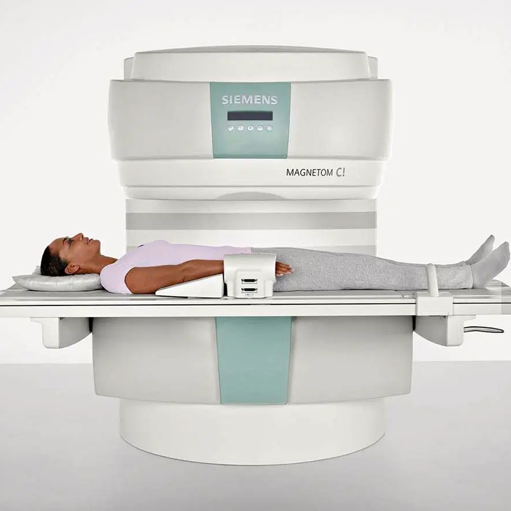
Wider and More Comfortable Scanning Area
One of the advantages of 0.35 Tesla (0.35T) MRI systems is their wider and more comfortable scanning area. Compared to traditional closed MRI machines, 0.35T MRI systems offer a more spacious and less confining environment for patients undergoing imaging procedures. This open design provides patients with a greater sense of comfort and reduces feelings of claustrophobia and anxiety during scanning.
The wider scanning area of 0.35T MRI systems also allows for easier positioning of patients, particularly those with larger body sizes or mobility issues. Patients have more room to maneuver and adjust themselves comfortably on the scanning table, improving their overall experience during the imaging procedure.
Additionally, the open design of 0.35T MRI systems can accommodate patients with special needs, such as those requiring assistance from caregivers or those with conditions that may make traditional closed MRI scanning challenging. This inclusivity ensures that a diverse range of patients can undergo MRI imaging comfortably and effectively.
Overall, the wider and more comfortable scanning area of 0.35T MRI systems enhances patient comfort and accessibility, contributing to a positive imaging experience for individuals undergoing MRI procedures.
The safety of 0.35 Tesla (0.35T) Open Magnetic Resonance Imaging (MRI) is a critical consideration in medical imaging. While MRI is generally considered safe, several factors contribute to ensuring the safety of patients undergoing MRI scans in 0.35T systems:
Magnetic Field Safety: The magnetic field strength of 0.35T MRI systems is lower compared to higher-field-strength MRI machines, which can contribute to reduced risks associated with magnetic field exposure. Patients with certain metallic implants or devices may be able to safely undergo imaging in 0.35T MRI systems, as the lower field strength poses less risk of heating or displacement of metallic objects.
Radiofrequency (RF) Safety: RF energy is used in MRI to generate the signals necessary for imaging. In 0.35T MRI systems, RF energy levels are optimized to minimize potential heating effects while still providing high-quality images. Safety measures such as patient screening for metallic implants and adherence to specific scanning protocols help ensure RF safety during imaging procedures.
Gradient Coil Safety: Gradient coils are used to create magnetic field gradients in MRI, enabling spatial encoding of the MRI signals. In 0.35T MRI systems, gradient coil designs prioritize patient safety by minimizing acoustic noise levels and adhering to safety guidelines for gradient field exposures.
Patient Monitoring: Continuous patient monitoring is essential during MRI scans to ensure patient safety and comfort. Medical staff closely monitor patients throughout the imaging procedure, addressing any concerns or adverse reactions promptly. Patient communication systems are also available to enable communication between patients and staff during the scan.
Safety Screening: Before undergoing an MRI scan in a 0.35T system, patients are screened for potential contraindications or safety concerns, such as the presence of metallic implants, pacemakers, or other medical devices that may pose risks during imaging. Clear communication between patients, radiologists, and other medical staff helps ensure that appropriate safety precautions are taken.
Overall, 0.35T Open MRI systems prioritize patient safety through optimized magnetic field, RF, and gradient coil safety measures, continuous patient monitoring, and thorough safety screening protocols. These measures contribute to the safe and effective utilization of MRI imaging in clinical practice.
Potential effects of the magnetic field in 0.35 Tesla (0.35T) Open Magnetic Resonance Imaging (MRI) systems and safety measures to mitigate risks include:
Magnetic Field Effects: The magnetic field generated by MRI systems can affect certain metallic objects or implants within the body. In 0.35T MRI systems, the lower magnetic field strength reduces the risk of adverse effects on metallic implants, such as heating or displacement. Safety measures include screening patients for metallic implants or devices before the scan and using specialized imaging protocols to minimize risks.
Heating of Metallic Implants: Metallic implants or devices within the body may experience heating when exposed to the magnetic field and radiofrequency (RF) energy during MRI scanning. In 0.35T MRI systems, the lower RF energy levels and optimized scanning protocols help minimize heating effects on metallic implants. Patients with metallic implants are carefully screened before the scan to assess the risk of heating and ensure their safety.
Implant Compatibility: Not all metallic implants are compatible with MRI scanning, regardless of the magnetic field strength. Safety measures include thorough patient screening to identify incompatible implants and using alternative imaging modalities when MRI is contraindicated. In cases where MRI is necessary, patients with compatible implants may undergo imaging safely in 0.35T MRI systems with appropriate precautions.
Acoustic Noise: Gradient coils used in MRI systems produce acoustic noise during scanning, which can be uncomfortable for patients. In 0.35T MRI systems, safety measures such as ear protection and noise-reduction techniques help minimize patient discomfort caused by acoustic noise.
Patient Monitoring: Continuous monitoring of patients during MRI scanning is essential to ensure their safety and well-being. Medical staff closely monitor patients for any signs of discomfort, adverse reactions, or physiological changes throughout the imaging procedure. Clear communication between patients and staff allows for prompt intervention if needed.
Overall, while 0.35T Open MRI systems offer advantages such as reduced magnetic field effects on metallic implants and improved patient comfort, safety measures such as thorough patient screening, optimized imaging protocols, and continuous patient monitoring are essential to mitigate potential risks and ensure the safe utilization of MRI imaging.
Indications and contraindications for the use of 0.35 Tesla (0.35T) Open Magnetic Resonance Imaging (MRI) systems include:
Indications:
- Neurological Imaging: Evaluation of brain tumors, strokes, multiple sclerosis, and other neurological conditions.
- Musculoskeletal Imaging: Assessment of joint injuries, ligament tears, osteoarthritis, and other musculoskeletal disorders.
- Spinal Imaging: Diagnosis of spinal cord injuries, herniated discs, spinal stenosis, and other spinal conditions.
- Abdominal Imaging: Evaluation of liver, kidney, and gastrointestinal disorders, as well as abdominal masses and organ abnormalities.
- Pediatric Imaging: Imaging of congenital anomalies, developmental disorders, and injuries in pediatric patients.
- Geriatric Imaging: Assessment of degenerative joint diseases, osteoporosis, and cerebrovascular diseases in elderly patients.
- Claustrophobic Patients: Alternative imaging option for patients with claustrophobia or anxiety disorders who may have difficulty tolerating traditional closed MRI scanners.
Contraindications:
- Metallic Implants: Presence of certain metallic implants or devices that are incompatible with MRI scanning, such as pacemakers, defibrillators, cochlear implants, and some orthopedic implants.
- Pregnancy: MRI scanning is generally avoided during the first trimester of pregnancy unless deemed necessary for diagnostic purposes and when the benefits outweigh the risks.
- Severe Claustrophobia: Patients with severe claustrophobia or anxiety disorders may still experience discomfort or distress in open MRI systems despite the more spacious environment.
- Inability to Cooperate: Patients who are unable to remain still or follow instructions during the imaging procedure may not be suitable candidates for MRI scanning, as motion artifacts can affect image quality.
- Acute Medical Conditions: Patients with acute medical conditions that require urgent medical intervention may not be suitable candidates for elective MRI scanning until their condition stabilizes.
It is essential for healthcare providers to carefully assess each patient’s medical history, clinical condition, and specific imaging needs to determine the appropriateness of using 0.35T Open MRI systems. Close communication between referring physicians, radiologists, and patients helps ensure safe and effective imaging practices.
Anatomical and pathological assessment abilities of 0.35 Tesla (0.35T) Open Magnetic Resonance Imaging (MRI) include:
Anatomical Detail: High-resolution imaging capabilities provide detailed visualization of anatomical structures, including organs, tissues, and anatomical landmarks, enabling precise anatomical assessment.
Tumor Detection and Characterization: MRI is sensitive to soft tissue contrast and can detect and characterize various types of tumors based on their size, location, morphology, and enhancement patterns, aiding in tumor staging and treatment planning.
Evaluation of Organ Morphology: MRI allows for the assessment of organ morphology and architecture, identifying abnormalities such as organ enlargement, atrophy, cysts, and structural deformities.
Detection of Inflammatory Changes: MRI can detect and characterize inflammatory changes in tissues, such as edema, inflammation, and vascular dilation, providing valuable information for diagnosing inflammatory conditions.
Assessment of Degenerative Changes: MRI is effective in assessing degenerative changes in musculoskeletal structures, including joint degeneration, cartilage loss, bone erosions, and disc degeneration, aiding in the diagnosis of degenerative joint diseases.
Identification of Vascular Anomalies: MRI can visualize vascular anomalies, such as aneurysms, vascular malformations, and stenoses, providing detailed anatomical information for surgical planning or endovascular interventions.
Evaluation of Neurological Pathologies: MRI is the imaging modality of choice for evaluating neurological pathologies, including brain tumors, stroke, multiple sclerosis, and neurodegenerative diseases, offering superior soft tissue contrast and multiplanar imaging capabilities.
Detection of Abdominal Abnormalities: MRI enables the detection and characterization of abdominal abnormalities, including liver lesions, pancreatic tumors, gastrointestinal pathology, and renal abnormalities, providing valuable diagnostic information in abdominal imaging.
Assessment of Spinal Pathologies: MRI is essential for evaluating spinal pathologies, including herniated discs, spinal cord compression, spinal tumors, and spinal stenosis, offering detailed visualization of the spinal cord, nerve roots, and surrounding structures.
Characterization of Soft Tissue Lesions: MRI is highly sensitive to soft tissue contrast and can characterize various soft tissue lesions, including cysts, abscesses, hematomas, and soft tissue tumors, aiding in differential diagnosis and treatment planning.
Overall, the anatomical and pathological assessment abilities of 0.35T MRI make it a valuable imaging tool for diagnosing and evaluating a wide range of medical conditions across different anatomical regions and organ systems.
High-quality imaging and diagnostic capability are key features of 0.35 Tesla (0.35T) Open Magnetic Resonance Imaging (MRI), offering several advantages:
Superior Soft Tissue Contrast: MRI utilizes powerful magnets and radiofrequency pulses to produce high-resolution images with excellent soft tissue contrast. This enables detailed visualization of anatomical structures, organs, and tissues, allowing for precise diagnostic assessment.
Multiplanar Imaging: 0.35T MRI systems provide multiplanar imaging capabilities, allowing for imaging in multiple planes (sagittal, axial, coronal), which is essential for comprehensive anatomical evaluation and precise localization of abnormalities.
Detailed Visualization: The high spatial resolution of 0.35T MRI enables detailed visualization of small anatomical structures and subtle pathological changes, facilitating accurate diagnosis and treatment planning.
Tumor Detection and Characterization: MRI is highly sensitive for detecting and characterizing tumors based on their size, location, morphology, and tissue characteristics. This includes the differentiation between benign and malignant lesions, aiding in tumor staging and treatment planning.
Functional Imaging Techniques: Advanced MRI techniques, such as diffusion-weighted imaging (DWI), perfusion-weighted imaging (PWI), and magnetic resonance spectroscopy (MRS), provide functional information about tissue microstructure, perfusion, and metabolism, enhancing diagnostic capability in oncology, neurology, and other fields.
Neurological Assessment: 0.35T MRI is indispensable for neurological imaging, allowing for detailed evaluation of the brain, spinal cord, and peripheral nerves. It aids in the diagnosis and management of various neurological disorders, including stroke, brain tumors, multiple sclerosis, and neurodegenerative diseases.
Musculoskeletal Evaluation: MRI offers comprehensive assessment of the musculoskeletal system, including bones, joints, muscles, and soft tissues. It is valuable for diagnosing orthopedic injuries, degenerative joint diseases, sports injuries, and musculoskeletal tumors.
Abdominal and Pelvic Imaging: 0.35T MRI provides high-quality imaging of abdominal and pelvic organs, allowing for the evaluation of liver, pancreas, kidneys, gastrointestinal tract, and reproductive organs. It aids in the diagnosis of abdominal masses, organ abnormalities, and gastrointestinal pathology.
Vascular Imaging: MRI offers non-invasive vascular imaging techniques, such as magnetic resonance angiography (MRA), for evaluating blood vessels throughout the body. It is useful for diagnosing vascular anomalies, stenoses, aneurysms, and arterial occlusions.
Comprehensive Diagnostic Capability: With its excellent soft tissue contrast, multiplanar imaging capabilities, and advanced functional imaging techniques, 0.35T MRI provides a comprehensive diagnostic tool for a wide range of medical conditions across various anatomical regions and organ systems.
Technological advancements in 0.35 Tesla (0.35T) Open Magnetic Resonance Imaging (MRI) have contributed to enhanced imaging capabilities and improved patient experience. Some key advancements include:
Improved Magnet Design: Advances in magnet design have led to more compact and efficient MRI systems, reducing space requirements and enhancing patient accessibility. Open MRI systems with a 0.35T magnetic field strength offer a more open and less confining environment compared to traditional closed-bore systems.
Higher Gradient Performance: Upgrades in gradient coil technology have resulted in higher gradient performance, enabling faster image acquisition and improved image quality. Higher gradient strength and slew rates enhance spatial resolution and reduce susceptibility artifacts, enhancing diagnostic accuracy.
Enhanced RF Coil Design: Innovations in radiofrequency (RF) coil design have improved signal-to-noise ratio (SNR) and image quality. Advanced RF coil arrays with multi-channel capabilities provide better coverage and sensitivity, allowing for more detailed anatomical and functional imaging.
Parallel Imaging Techniques: Parallel imaging techniques, such as sensitivity encoding (SENSE) and generalized autocalibrating partially parallel acquisitions (GRAPPA), have been integrated into 0.35T MRI systems. These techniques accelerate image acquisition by simultaneously acquiring multiple spatially encoded signals, reducing scan time and motion artifacts.
Advanced Pulse Sequences: Development of advanced pulse sequences, including turbo spin-echo (TSE), fast spin-echo (FSE), and gradient echo (GRE) sequences, enables rapid imaging with improved contrast and resolution. Techniques like diffusion-weighted imaging (DWI) and magnetic resonance spectroscopy (MRS) provide functional and metabolic information, enhancing diagnostic capabilities.
Motion Correction and Artifact Reduction: Motion correction techniques, such as prospective motion correction and motion-sensitive pulse sequences, mitigate motion artifacts caused by patient movement. Advanced reconstruction algorithms and post-processing techniques further reduce artifacts and improve image quality.
Real-time Imaging: Real-time imaging capabilities allow for dynamic visualization of physiological processes and anatomical structures in motion. Real-time MRI techniques, such as cine imaging and dynamic contrast-enhanced (DCE) MRI, enable functional assessment of cardiac function, joint dynamics, and gastrointestinal motility.
Integrated Workflow Solutions: Integration of MRI systems with advanced workflow solutions, including automated image reconstruction, post-processing software, and image analysis tools, streamlines workflow and enhances diagnostic efficiency. Automated image registration and fusion techniques enable multi-modality image integration for comprehensive diagnosis.
Patient Comfort and Experience: Technological advancements in patient comfort features, such as quieter gradient coils, reduced acoustic noise, and improved bore design, enhance the overall patient experience during MRI examinations. Open MRI systems with wider bores and shorter scan times reduce claustrophobia and anxiety, improving patient compliance and satisfaction.
Accessibility and Affordability: Advances in MRI technology have led to the development of more affordable and accessible 0.35T MRI systems, expanding access to high-quality imaging in diverse clinical settings. These advancements contribute to improved patient care and diagnostic accuracy across various medical specialties.
Advancements in imaging techniques have revolutionized diagnostic capabilities in various medical fields. Some key advancements include:
Multi-Parametric Imaging: Integrating multiple imaging parameters, such as T1-weighted, T2-weighted, diffusion-weighted, and dynamic contrast-enhanced sequences, allows for comprehensive characterization of tissue morphology, vascularity, and functional information within a single examination.
Functional Imaging: Functional imaging techniques, including functional MRI (fMRI), diffusion tensor imaging (DTI), and magnetic resonance spectroscopy (MRS), enable the assessment of tissue function and metabolism. These techniques provide valuable insights into brain function, tumor biology, and metabolic abnormalities.
Quantitative Imaging: Quantitative imaging techniques, such as diffusion kurtosis imaging (DKI), arterial spin labeling (ASL), and magnetic resonance elastography (MRE), provide quantitative measurements of tissue properties, such as diffusivity, perfusion, and stiffness. These techniques offer valuable biomarkers for disease diagnosis, monitoring, and treatment response assessment.
Spectroscopic Imaging: Magnetic resonance spectroscopic imaging (MRSI) enables the non-invasive assessment of tissue metabolites, such as choline, creatine, and N-acetylaspartate, providing insights into tissue biochemistry and metabolic abnormalities. MRSI is valuable for investigating various neurological, oncological, and metabolic disorders.
Diffusion Weighted Imaging (DWI): DWI measures the diffusion of water molecules within tissues, offering insights into tissue microstructure and cellular integrity. DWI is widely used for diagnosing acute ischemic stroke, characterizing tumors, and assessing tissue viability in various organs.
Perfusion Imaging: Dynamic contrast-enhanced MRI (DCE-MRI) and arterial spin labeling (ASL) techniques enable the assessment of tissue perfusion and blood flow. Perfusion imaging is valuable for evaluating tissue vascularity, tumor angiogenesis, and vascular disorders.
Functional Connectivity Mapping: Resting-state fMRI (rs-fMRI) and diffusion tensor imaging (DTI) enable the mapping of functional connectivity within the brain and white matter tracts, respectively. Functional connectivity mapping provides insights into brain networks, neuroplasticity, and neurological disorders.
Ultra-High Field MRI: Advancements in ultra-high field (7T and above) MRI systems offer increased spatial resolution, signal-to-noise ratio (SNR), and spectral dispersion. Ultra-high field MRI enables detailed visualization of small anatomical structures, cellular-level imaging, and advanced spectroscopic techniques.
Artificial Intelligence (AI) in Imaging: AI-driven imaging techniques, including machine learning, deep learning, and computer-aided diagnosis (CAD), enhance image reconstruction, segmentation, and quantitative analysis. AI algorithms improve diagnostic accuracy, reduce interpretation time, and facilitate personalized treatment planning.
Hybrid Imaging Modalities: Integration of MRI with other imaging modalities, such as positron emission tomography (PET), single-photon emission computed tomography (SPECT), and computed tomography (CT), enables hybrid imaging techniques. Hybrid imaging offers complementary functional and anatomical information, improving diagnostic accuracy in oncology, cardiology, and neurology.
These advancements in imaging techniques continue to drive innovation in medical imaging, enabling precise diagnosis, personalized treatment planning, and improved patient outcomes across a wide range of medical specialties.
Accessibility and availability of medical imaging services, including Magnetic Resonance Imaging (MRI), are essential for ensuring timely diagnosis and treatment for patients. Several factors contribute to enhancing accessibility and availability:
Geographical Distribution: Establishing MRI facilities in strategically located areas ensures geographic accessibility for patients across urban, suburban, and rural regions. Equitably distributing MRI centers reduces travel distances and improves access for individuals living in remote or underserved areas.
Multi-Modality Imaging Centers: Integrating MRI services within multi-modality imaging centers that offer a comprehensive range of diagnostic imaging modalities, such as X-ray, computed tomography (CT), ultrasound, and nuclear medicine, enhances accessibility for patients requiring diverse imaging studies.
Mobile MRI Units: Deploying mobile MRI units equipped with advanced imaging technology enables on-site imaging services at hospitals, clinics, and remote healthcare facilities. Mobile MRI units are particularly beneficial for serving patients with limited mobility, those in long-term care facilities, or during emergency situations.
Extended Operating Hours: Extending MRI operating hours beyond regular business hours, including evenings and weekends, accommodates patients with scheduling constraints due to work, school, or other commitments. Flexible scheduling options improve accessibility and reduce wait times for imaging appointments.
Appointment Scheduling Systems: Implementing efficient appointment scheduling systems, including online booking platforms and centralized scheduling centers, streamlines the appointment process and reduces wait times for patients. Offering same-day or next-day appointments for urgent cases enhances accessibility for patients requiring expedited imaging services.
Referral Pathways: Establishing clear referral pathways and communication channels between healthcare providers, primary care physicians, specialists, and imaging centers facilitates timely access to MRI services. Efficient referral processes ensure that patients receive appropriate imaging studies based on their clinical needs.
Patient Education and Outreach: Conducting patient education and outreach programs to raise awareness about the importance of medical imaging, including MRI, and its role in disease diagnosis and management encourages proactive healthcare-seeking behavior. Educating patients about the availability of MRI services and addressing common concerns or misconceptions enhances accessibility.
Insurance Coverage and Reimbursement: Ensuring adequate insurance coverage and reimbursement for MRI services through public and private health insurance programs increases affordability and accessibility for patients. Transparent billing practices and financial assistance programs further support patients in accessing MRI services.
By implementing these strategies, healthcare providers and imaging centers can improve the accessibility and availability of MRI services, ultimately enhancing patient care and outcomes.
The future of Open Magnetic Resonance Imaging (MRI) holds promising advancements and innovations aimed at further enhancing imaging capabilities, patient experience, and clinical utility. Some key aspects of the future of Open MRI include:
Advanced Imaging Techniques: Ongoing research and development efforts focus on refining imaging techniques to improve spatial and temporal resolution, tissue contrast, and functional imaging capabilities. Advancements in multi-parametric imaging, quantitative imaging, and spectroscopic techniques will enable more comprehensive and accurate diagnosis of various medical conditions.
Ultra-High Field Open MRI: Technological advancements may lead to the development of ultra-high field Open MRI systems with magnetic field strengths exceeding 1.5 Tesla. These systems offer increased signal-to-noise ratio (SNR) and spatial resolution, enabling detailed visualization of anatomical structures and pathological processes.
Functional and Molecular Imaging: Future Open MRI systems may incorporate advanced functional imaging techniques, such as functional MRI (fMRI), diffusion tensor imaging (DTI), and magnetic resonance spectroscopy (MRS), to assess tissue function and metabolism. Molecular imaging modalities, including targeted contrast agents and molecular probes, will facilitate non-invasive detection and characterization of molecular biomarkers associated with disease.
Artificial Intelligence (AI) Integration: Integration of AI-driven algorithms and machine learning techniques into Open MRI systems will optimize image acquisition, reconstruction, and analysis. AI algorithms will enhance image quality, automate image interpretation, and provide quantitative assessment of imaging biomarkers, improving diagnostic accuracy and efficiency.
Personalized Imaging Protocols: Future Open MRI systems may feature personalized imaging protocols tailored to individual patient characteristics, such as age, body habitus, and specific clinical indications. Adaptive imaging techniques and real-time feedback mechanisms will optimize imaging parameters to achieve optimal diagnostic performance while minimizing scan time and patient discomfort.
Patient-Centered Design: Continued focus on patient-centered design principles will drive the development of Open MRI systems that prioritize patient comfort, accessibility, and safety. Innovations in bore design, noise reduction technology, and motion correction techniques will enhance the overall patient experience during MRI examinations.
Integrated Multimodal Imaging: Integration of Open MRI with other imaging modalities, such as positron emission tomography (PET), computed tomography (CT), and ultrasound, will enable multimodal imaging approaches for comprehensive disease evaluation. Hybrid imaging systems combining multiple modalities within a single platform will facilitate complementary anatomical and functional imaging, enhancing diagnostic accuracy and treatment planning.
Remote Monitoring and Telemedicine: Advancements in remote monitoring technologies and telemedicine platforms will enable remote interpretation of Open MRI images and consultations with radiologists. Tele-radiology services will improve access to expert interpretation in underserved regions and facilitate collaboration among healthcare providers for multidisciplinary patient care.
Overall, the future of Open MRI holds immense potential for advancing medical imaging capabilities, transforming patient care, and driving innovation in diagnostic radiology. Continued research and collaboration among industry stakeholders will play a crucial role in realizing these advancements and shaping the future landscape of Open MRI technology.
Evolution and Changing Role in Clinical Applications
The evolution of Open Magnetic Resonance Imaging (MRI) technology has significantly impacted its role in clinical applications. As MRI systems have advanced, they have become indispensable tools in various medical specialties, ranging from neurology and orthopedics to oncology and cardiology.
Future Directions and Potential Improvements
Looking ahead, ongoing research and development efforts aim to further enhance Open MRI technology to meet evolving clinical needs. Innovations in imaging techniques, hardware design, and software algorithms promise to improve image quality, diagnostic accuracy, and patient comfort.
Frequently Asked Questions
Get a Free Second Opinion
Experienced Burtom Medical Team is Ready to Help
I consent to Burtom Health Group using my aforesaid personal data for the purposes described in this notice and understand that I can withdraw my consent at any time by sending a request to info@burtom.com.


