Burtom ‣ Technologies ‣ Podoscope (Foot Graph)
Language: 🇬🇧 English | 🇹🇷 Türkçe
Podoscope (Foot Graph) Overview

 A podoscope, commonly referred to as a foot graph, is a specialized medical device used in podiatry and orthopedics to assess the structure and function of the feet. It consists of a platform equipped with sensors or a pressure-sensitive mat that captures data related to foot pressure distribution. This information is then visualized on a computer screen or printed out as a foot pressure map, allowing healthcare professionals to analyze various aspects of foot mechanics and gait.
One of the primary functions of a podoscope is to aid in the diagnosis and treatment of foot-related conditions such as plantar fasciitis, flat feet, and diabetic foot ulcers. By assessing the pressure distribution under the feet, podiatrists can identify areas of high pressure, abnormal gait patterns, and potential sources of discomfort or injury. This information guides treatment planning, which may include orthotic interventions, footwear modifications, or therapeutic exercises aimed at improving foot function and relieving pain.
Furthermore, podoscopes play a crucial role in biomechanical research and sports medicine. Researchers use these devices to study the effects of different activities, footwear designs, and orthotic interventions on foot mechanics and performance. Athletes, particularly runners and dancers, benefit from podoscopic assessments to optimize their gait patterns, prevent injuries, and enhance overall athletic performance. Overall, podoscopes are valuable tools in both clinical practice and research settings for assessing foot health, diagnosing conditions, and improving treatment outcomes.


What is Podoscope (Foot Graph) and How Does It Work?
Podoscope, a medical device used to examine and analyze foot structure. This device is designed to visualize the sole of the foot in detail and evaluate various conditions related to foot health. The working principle of the podoscope involves using a special optical system and surface detection technology to measure pressure points on the sole and examine the foot anatomy in detail. This allows for the detection of issues such as asymmetry, deformities, or pressure points in the foot structure, providing valuable information for treatment planning. The podoscope serves as an important tool in evaluating foot health, aiding in the early diagnosis of various foot-related conditions and monitoring treatment processes.
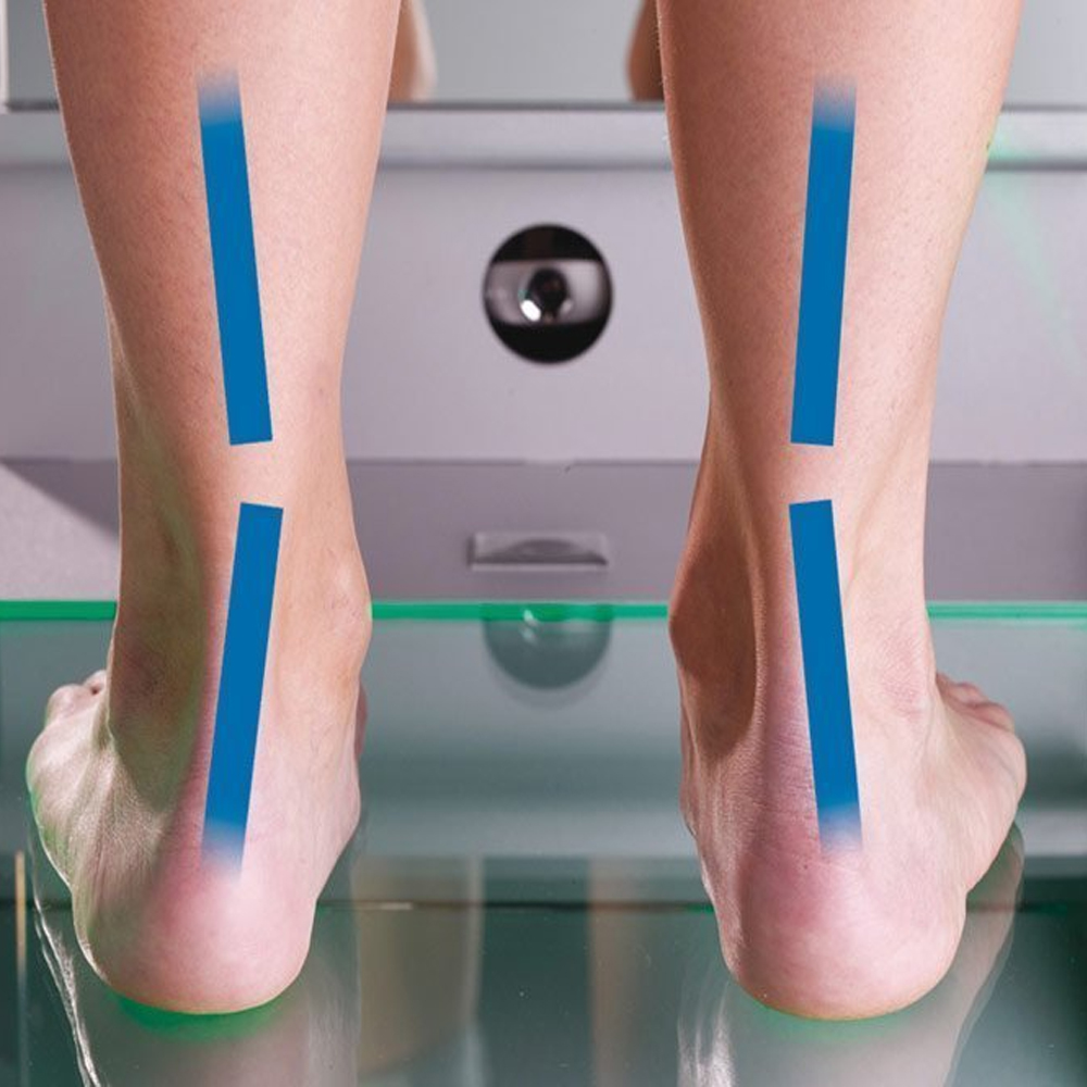
Basic Principles and Principles of Podoscope (Foot Graph): The podoscope, also known as a foot graph, operates based on several basic principles and principles. It utilizes specialized optical systems and surface detection technology to visualize and analyze the structure of the foot. By capturing detailed images and pressure distributions on the sole of the foot, the podoscope provides valuable insights into foot anatomy, identifying areas of asymmetry, deformities, or abnormal pressure points.
How is the Structure of the Foot Visualized and Analyzed?: The visualization and analysis of the foot structure are achieved through the use of advanced imaging techniques and pressure-sensitive sensors integrated into the podoscope. These sensors detect and measure pressure distribution across the sole of the foot, allowing for a comprehensive assessment of foot health.
Applications and Importance of Podoscope (Foot Graph): The podoscope plays a crucial role in various applications related to foot health. It is commonly used in orthopedic evaluations and treatment planning to assess foot conditions such as flat feet, high arches, bunions, and other structural abnormalities. Additionally, podoscopes are utilized in podiatry clinics, sports medicine facilities, and footwear research laboratories to analyze gait patterns, evaluate biomechanical function, and design customized orthotic devices. Overall, the podoscope serves as a valuable tool in podiatric medicine, providing clinicians with essential information for diagnosing foot disorders, optimizing treatment strategies, and improving patient outcomes.
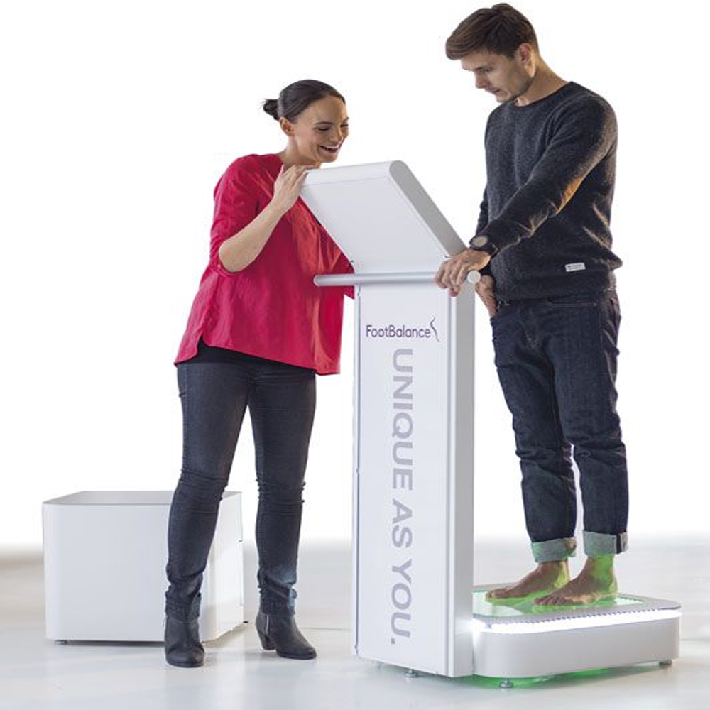
Podoscope System and Basic Components: The podoscope system consists of several essential components designed to facilitate accurate foot analysis. These components typically include a durable platform with pressure-sensitive sensors, an integrated camera system for capturing foot images, and specialized software for data processing and analysis. Additionally, the system may feature adjustable lighting options and ergonomic design elements to ensure patient comfort and ease of use.
Image Acquisition and Processing: Image acquisition and processing are integral parts of the podoscope system, enabling clinicians to obtain detailed visualizations of the foot’s structure and pressure distribution. The integrated camera captures high-resolution images of the foot while the pressure-sensitive sensors record pressure points and weight distribution. These data are then processed and analyzed using specialized software, allowing clinicians to assess foot biomechanics, identify abnormalities, and tailor treatment plans accordingly.
Clinical Applications of Podoscope (Foot Graph): The podoscope, or foot graph, finds widespread application across various clinical settings and specialties. In orthopedics, it is used to evaluate foot deformities, such as pes planus (flat feet) or pes cavus (high arches), and assess the effectiveness of orthotic interventions. Podiatrists utilize podoscopes to diagnose and monitor conditions like plantar fasciitis, Morton’s neuroma, and diabetic foot ulcers, enabling early intervention and prevention of complications. Moreover, sports medicine professionals utilize podoscopes to analyze gait patterns, assess biomechanical imbalances, and optimize athletic performance. Overall, the podoscope serves as a valuable diagnostic tool in podiatric medicine, providing clinicians with valuable insights into foot health and function.
Preoperative Planning for Orthopedic Surgery: Podoscope (Foot Graph) plays a crucial role in preoperative planning for orthopedic surgery, especially procedures involving the foot and ankle. By providing detailed visualizations of foot structure and pressure distribution, podoscopy aids orthopedic surgeons in developing precise surgical plans. Surgeons can assess biomechanical abnormalities, identify areas of high pressure or stress, and determine the optimal surgical approach to address specific conditions, such as bunions, hammertoes, or structural deformities.
Evaluation of Injuries and Disorders Occurring in the Foot: Podoscope (Foot Graph) serves as a valuable diagnostic tool for evaluating injuries and disorders affecting the foot. Podiatric physicians and orthopedic specialists utilize podoscopy to assess various foot conditions, including plantar fasciitis, Achilles tendonitis, stress fractures, and neuropathic ulcers. By analyzing pressure distribution patterns and foot biomechanics, clinicians can accurately diagnose these conditions, monitor disease progression, and tailor treatment strategies accordingly. Additionally, podoscopy aids in identifying contributing factors to foot injuries, such as improper footwear or gait abnormalities, facilitating comprehensive treatment planning.
Advantages and Benefits of Podoscope (Foot Graph): The utilization of podoscopy offers numerous advantages and benefits in clinical practice. Firstly, podoscopy provides detailed visualizations of foot anatomy and pressure distribution, enabling precise diagnosis and treatment planning for various foot conditions. Additionally, podoscopy facilitates early detection of foot abnormalities, allowing for proactive intervention and prevention of complications. Furthermore, podoscopy supports patient education by visually demonstrating the impact of foot pathology on gait and weight-bearing, empowering patients to actively participate in their treatment plans. Overall, the integration of podoscopy into clinical practice enhances diagnostic accuracy, improves treatment outcomes, and enhances the overall quality of foot care.
High Resolution and Detailed Imaging: Podoscope (Foot Graph) offers high-resolution and detailed imaging capabilities, providing healthcare professionals with clear visualizations of foot anatomy. By capturing precise images of the foot’s plantar surface, including pressure distribution patterns and weight-bearing areas, podoscopy enables accurate assessment of foot structure and biomechanics. This level of detail allows clinicians to identify subtle abnormalities, such as pressure points, deformities, or gait abnormalities, that may contribute to foot-related conditions or injuries.
Precise Examination and Measurements of Foot Anatomy: One of the key advantages of podoscopy is its ability to facilitate precise examination and measurements of foot anatomy. Through advanced imaging technology and specialized software, podoscopy allows clinicians to conduct detailed analyses of foot morphology, including arch height, foot length, and width. Additionally, podoscopy enables the assessment of dynamic parameters such as gait dynamics and pressure distribution during walking or running. These precise measurements aid in the diagnosis of various foot conditions, the customization of orthotic devices, and the monitoring of treatment outcomes over time.
Safe Use of Podoscope (Foot Graph): Safety is paramount when utilizing podoscopy in clinical practice. Healthcare providers must adhere to established safety protocols and guidelines to ensure the safe use of podoscope systems. This includes proper calibration and maintenance of equipment to ensure accurate imaging and reliable performance. Additionally, clinicians should undergo comprehensive training to operate podoscopy systems safely and effectively, minimizing the risk of errors or injuries during examinations. Furthermore, patient safety measures, such as ensuring proper positioning and comfort during imaging procedures, are essential to optimize the patient experience and minimize discomfort or complications. Overall, prioritizing safety measures ensures that podoscopy remains a valuable and safe tool for foot assessment in clinical settings.
Radiation Exposure and Safety Measures: Podoscope (Foot Graph) imaging typically does not involve ionizing radiation, as it primarily relies on optical or digital technologies to capture foot anatomy. Therefore, the risk of radiation exposure associated with podoscopy is minimal compared to traditional X-ray imaging modalities. However, if podoscopy systems incorporate any radiation-emitting components, such as fluoroscopy for dynamic foot assessments, appropriate safety measures must be implemented to minimize radiation exposure. This includes adhering to regulatory guidelines, such as ALARA (As Low As Reasonably Achievable) principles, and ensuring that healthcare professionals and patients are adequately shielded from unnecessary radiation.
Precautions for Patient Safety During Podoscope Procedure: Ensuring patient safety is essential during podoscopy procedures. Healthcare providers must take several precautions to minimize the risk of injury or discomfort for patients. This includes verifying patient suitability for podoscopy, considering factors such as medical history, allergies, and contraindications to foot manipulation or pressure. Proper patient positioning and stabilization are crucial to obtain accurate and reproducible foot images while ensuring patient comfort throughout the procedure. Additionally, clinicians should communicate clearly with patients, explaining the purpose of the examination, addressing any concerns or questions, and obtaining informed consent before proceeding with the podoscopy procedure.
Evaluation and Interpretation of Podoscope Images: Following podoscopy imaging, healthcare professionals must accurately evaluate and interpret the obtained images to derive meaningful clinical insights. This involves a thorough analysis of foot morphology, pressure distribution patterns, and biomechanical parameters captured during the examination. Specialized software tools may aid in image analysis, allowing for precise measurements of foot dimensions, arch height, pressure points, and gait dynamics. Clinicians should integrate this quantitative data with qualitative observations to formulate comprehensive assessments of foot health and function. Clear communication of findings and interpretation with patients and interdisciplinary healthcare teams facilitates collaborative decision-making and the development of tailored treatment plans for foot-related conditions or injuries.
Understanding Image Results and Sharing with the Doctor: After capturing foot images using a podoscope (foot graph), it is essential to interpret the results accurately to understand foot anatomy and pathology. Healthcare professionals must carefully analyze the images to identify any abnormalities or irregularities in foot structure, such as arch height, pressure distribution patterns, or deformities. Clear communication and collaboration with physicians and specialists are crucial to ensure accurate diagnosis and effective treatment planning. By sharing detailed image results with the doctor, clinicians can facilitate interdisciplinary discussions, enabling comprehensive assessments and informed decision-making regarding foot health and management strategies.
Use and Monitoring of Images in the Treatment Process: Images obtained through podoscopy play a vital role in the treatment process, serving as valuable diagnostic tools and guiding therapeutic interventions for various foot-related conditions. Healthcare providers utilize these images to monitor changes in foot morphology, assess treatment efficacy, and track patient progress over time. By comparing sequential podoscope images, clinicians can evaluate the effectiveness of interventions such as orthotic interventions, physical therapy, or surgical procedures. Regular monitoring of foot images enables healthcare professionals to adjust treatment plans as needed, ensuring optimal outcomes and improved patient satisfaction.
Improvements in Podoscope Technology and Future Potential: Advancements in podoscope technology continue to enhance foot imaging capabilities, offering opportunities for improved diagnostic accuracy and clinical outcomes. Innovations in imaging sensors, optics, and software algorithms enable higher resolution and more detailed visualization of foot anatomy and biomechanics. Furthermore, integration with digital health platforms and telemedicine applications facilitates remote image acquisition, analysis, and consultation, expanding access to podoscopy services for patients in diverse settings. The future potential of podoscope technology includes the development of artificial intelligence-driven image analysis tools, personalized treatment algorithms, and predictive models for foot health outcomes. These advancements hold promise for advancing podoscopic diagnostic capabilities, optimizing treatment strategies, and enhancing patient care in podiatry and orthopedics.
Patient Rights and Information About Podoscope (Foot Graph): Patients undergoing podoscopy have the right to receive comprehensive information about the procedure, including its purpose, potential benefits, risks, and alternatives. Healthcare providers should ensure that patients understand the nature of podoscopy, how foot images will be captured and used, and any associated costs. Patients should also be informed about their rights regarding the use and storage of their podoscope images, including consent requirements for sharing data with other healthcare professionals or third-party service providers. Clear communication and transparency regarding patient rights and information are essential to promoting patient autonomy and facilitating informed decision-making throughout the podoscopy process.
Privacy and Data Protection: Privacy and data protection are paramount considerations in podoscopy to safeguard patient confidentiality and comply with legal and ethical standards. Healthcare providers must implement robust data protection measures to secure podoscope images and associated patient information against unauthorized access, disclosure, or misuse. This includes encryption of electronic data, restricted access controls, and adherence to applicable privacy regulations such as the Health Insurance Portability and Accountability Act (HIPAA) in the United States or the General Data Protection Regulation (GDPR) in the European Union. Additionally, healthcare facilities should have clear policies and procedures in place for data retention, disposal, and breach response to mitigate privacy risks and ensure compliance with privacy laws.
Patient Rights and Preferences During the Procedure: Respecting patient rights and preferences is integral to delivering patient-centered care during podoscopy procedures. Healthcare providers should prioritize patient comfort and well-being throughout the examination, addressing any concerns or questions raised by the patient. Patients have the right to consent or refuse podoscopy based on their preferences, and healthcare providers must respect their autonomy in decision-making. Moreover, clinicians should accommodate patient preferences regarding factors such as the timing of the procedure, choice of imaging equipment, and involvement of support persons during the examination. By prioritizing patient rights and preferences, healthcare providers can enhance the patient experience and promote positive outcomes in podiatric care.
Future Applications and Development of Podoscope (Foot Graph): As technology continues to advance, the future of podoscopy holds promising opportunities for further innovation and development. Future applications of podoscope systems may include enhanced imaging techniques to provide even more detailed and comprehensive analysis of foot anatomy and function. Additionally, advancements in artificial intelligence (AI) and machine learning algorithms could enable automated interpretation of podoscope images, facilitating quicker diagnosis and treatment planning. Furthermore, integration with other digital health technologies, such as telemedicine platforms and wearable devices, may expand the reach of podoscopy beyond traditional clinical settings, allowing for remote monitoring and personalized interventions. Continued research and collaboration among healthcare professionals, engineers, and researchers are essential to unlocking the full potential of podoscopy and its future applications in podiatric care.
New Technologies and Application Areas: Emerging technologies hold the potential to revolutionize podoscopy and open up new application areas in foot health management. For example, advancements in 3D printing and personalized orthotics could be integrated with podoscope systems to create custom-fit footwear solutions tailored to individual foot morphology and biomechanics. Moreover, the integration of augmented reality (AR) or virtual reality (VR) technologies with podoscopy could enhance patient education and rehabilitation by providing immersive experiences and interactive visualizations of foot conditions and treatment options. Additionally, the development of miniaturized and portable podoscope devices could facilitate point-of-care diagnostics in various settings, including sports medicine, diabetic foot care, and geriatric care. By harnessing these new technologies, podoscopy can expand its reach and impact across diverse application areas in foot health management.
Development and Improvement Potential of Podoscope Systems: The ongoing development and improvement of podoscope systems are crucial for enhancing their effectiveness, usability, and clinical utility. Future efforts may focus on optimizing imaging modalities and sensor technologies to achieve higher resolution and more accurate foot measurements. Additionally, advancements in ergonomic design and user interface features could improve the ergonomics and user experience of podoscope devices, making them more user-friendly for healthcare providers and comfortable for patients. Furthermore, research into novel imaging techniques, such as hyperspectral imaging or multispectral imaging, may enable the detection of subtle changes in foot tissue composition and vascularity, enhancing diagnostic capabilities in podiatric practice. Collaborative efforts between industry stakeholders, researchers, and healthcare professionals will be essential to drive the continued development and improvement of podoscope systems, ultimately benefiting patients and advancing foot health care.
Frequently Asked Questions
Get a Free Second Opinion
Experienced Burtom Medical Team is Ready to Help
I consent to Burtom Health Group using my aforesaid personal data for the purposes described in this notice and understand that I can withdraw my consent at any time by sending a request to info@burtom.com.


